Deposition Date
2013-06-12
Release Date
2014-09-10
Last Version Date
2024-02-28
Entry Detail
PDB ID:
4L6C
Keywords:
Title:
Crystal structure of human mitochondrial deoxyribonucleotidase in complex with the inhibitor pib-t
Biological Source:
Source Organism(s):
Homo sapiens (Taxon ID: 9606)
Expression System(s):
Method Details:
Experimental Method:
Resolution:
1.80 Å
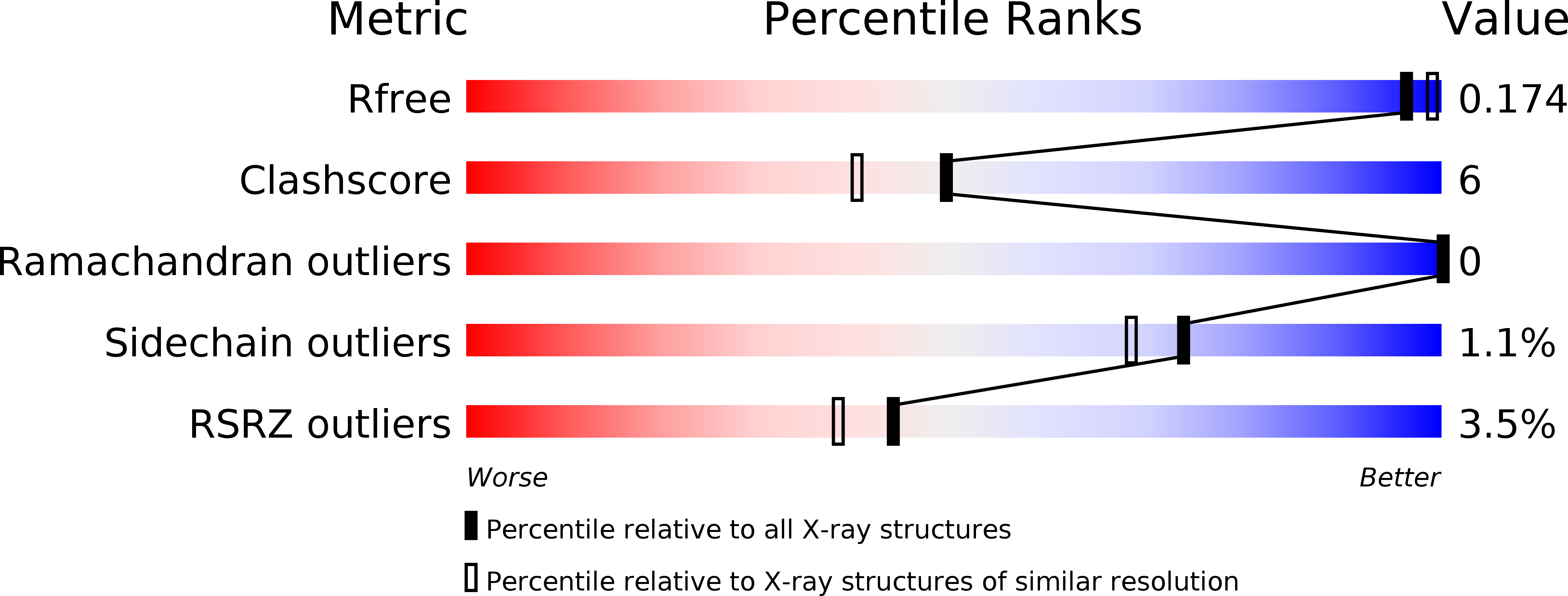
R-Value Free:
0.17
R-Value Work:
0.15
R-Value Observed:
0.15
Space Group:
P 43 21 2