
Deposition Date
2013-06-07
Release Date
2013-08-28
Last Version Date
2024-11-27
Entry Detail
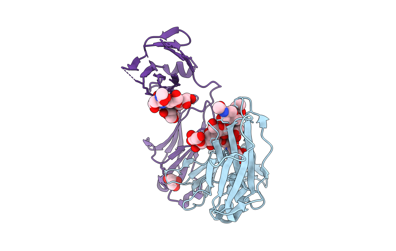
PDB ID:
4L4J
Keywords:
Title:
Crystal structure of fc-fragment of human IgG2-Sigma antibody
Biological Source:
Source Organism(s):
Homo sapiens (Taxon ID: 9606)
Expression System(s):
Method Details:
Experimental Method:
Resolution:
1.92 Å
R-Value Free:
0.24
R-Value Work:
0.19
R-Value Observed:
0.20
Space Group:
P 21 21 21
