Deposition Date
2013-06-05
Release Date
2013-12-04
Last Version Date
2024-11-20
Entry Detail
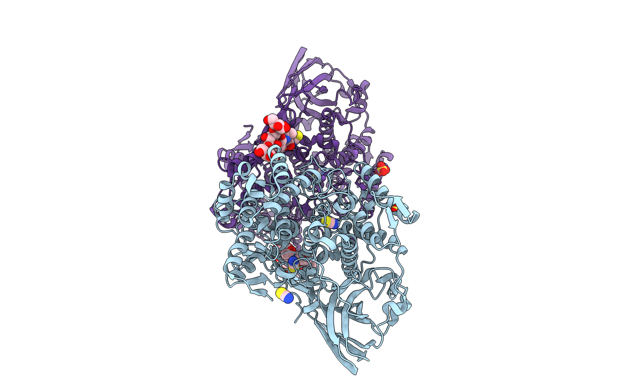
PDB ID:
4L37
Keywords:
Title:
SP2-SP3 - a complex of two storage proteins from Bombyx mori hemolymph
Biological Source:
Source Organism(s):
Bombyx mori (Taxon ID: 7091)
Method Details:
Experimental Method:
Resolution:
2.90 Å
R-Value Free:
0.22
R-Value Work:
0.16
R-Value Observed:
0.16
Space Group:
P 63 2 2