4L2M image
Deposition Date 2013-06-04
Release Date 2013-06-12
Last Version Date 2024-11-20
Entry Detail
PDB ID:
4L2M
Title:
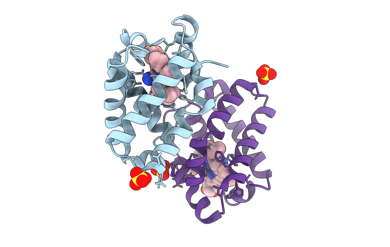
Crystal structure of the 2/2 hemoglobin from Synechococcus sp. PCC 7002 in the cyanomet state and with covalently attached heme
Biological Source:
Source Organism:
Host Organism:
Method Details:
Experimental Method:
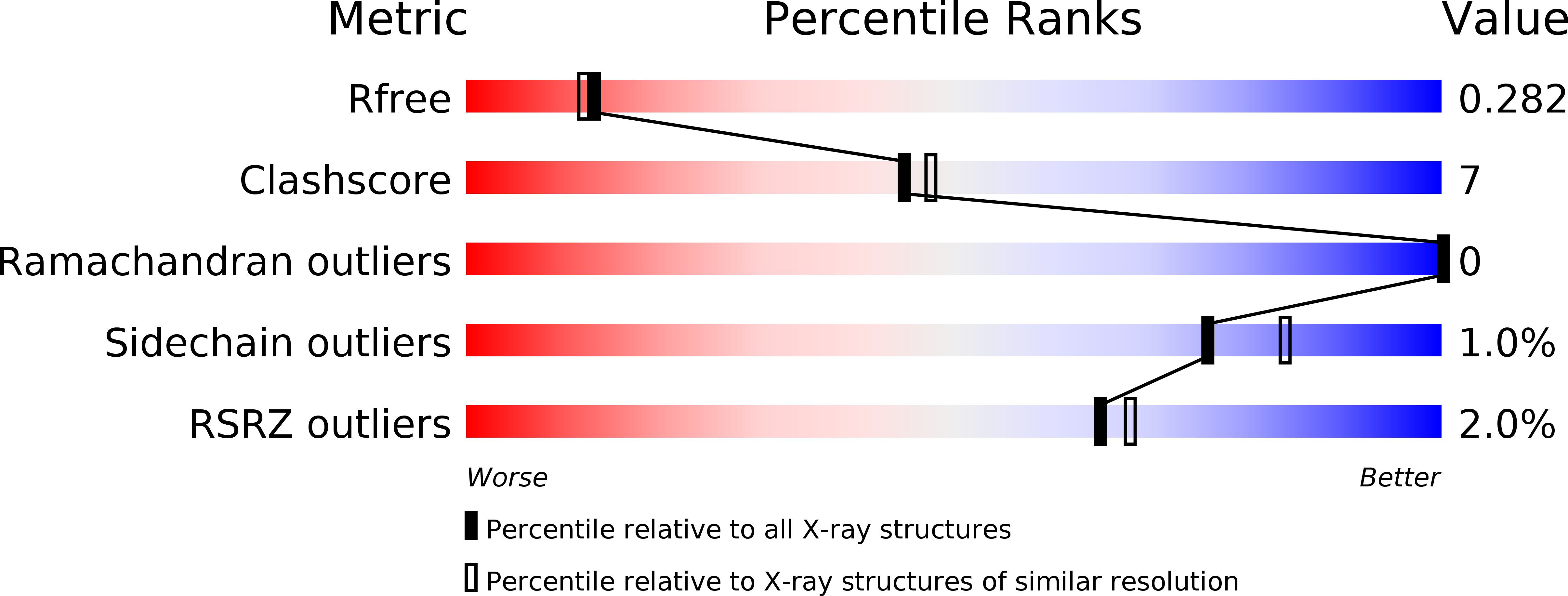
Resolution:
2.25 Å
R-Value Free:
0.28
R-Value Work:
0.21
R-Value Observed:
0.22
Space Group:
P 1 21 1
Macromolecular Entities
Polymer Type:polypeptide(L)
Molecule:Cyanoglobin
Gene (Uniprot):glbN
Chain IDs:A, B
Chain Length:123
Number of Molecules:2
Biological Source:Synechococcus sp.
Primary Citation
The 2/2 hemoglobin from the cyanobacterium Synechococcus sp. PCC 7002 with covalently attached heme: Comparison of X-ray and NMR structures.
Proteins 82 528 534 (2014)
PMID: 23999883 DOI: 10.1002/prot.24409

Abstact

The X-ray structures of the hemoglobin from Synechococcus sp. PCC 7002 (GlbN) were solved in the ferric bis-histidine (1.44 Å resolution) and cyanide-bound (2.25 Å resolution) states with covalently attached heme. The two structures illustrate the conformational changes and cavity opening caused by exogenous ligand binding. They also reveal an unusually distorted heme, ruffled as in c cytochromes. Comparison to the solution structure demonstrates the influence of crystal packing on several structural elements, whereas comparison to GlbN from Synechocystis sp. PCC 6803 shows subtle differences in heme geometries and environment. The new structures will be instrumental in elucidating GlbN reactivity.

Legend

Protein

Chemical

Disease

Primary Citation of related structures