Deposition Date
2013-06-03
Release Date
2013-10-23
Last Version Date
2024-02-28
Entry Detail
PDB ID:
4L1C
Keywords:
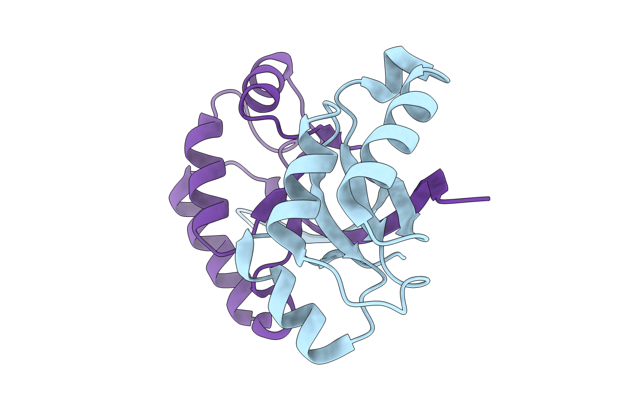
Title:
Crystal structure of Dimerized N-terminal Domain of MinC
Biological Source:
Source Organism(s):
Escherichia coli (Taxon ID: 364106)
Expression System(s):
Method Details:
Experimental Method:
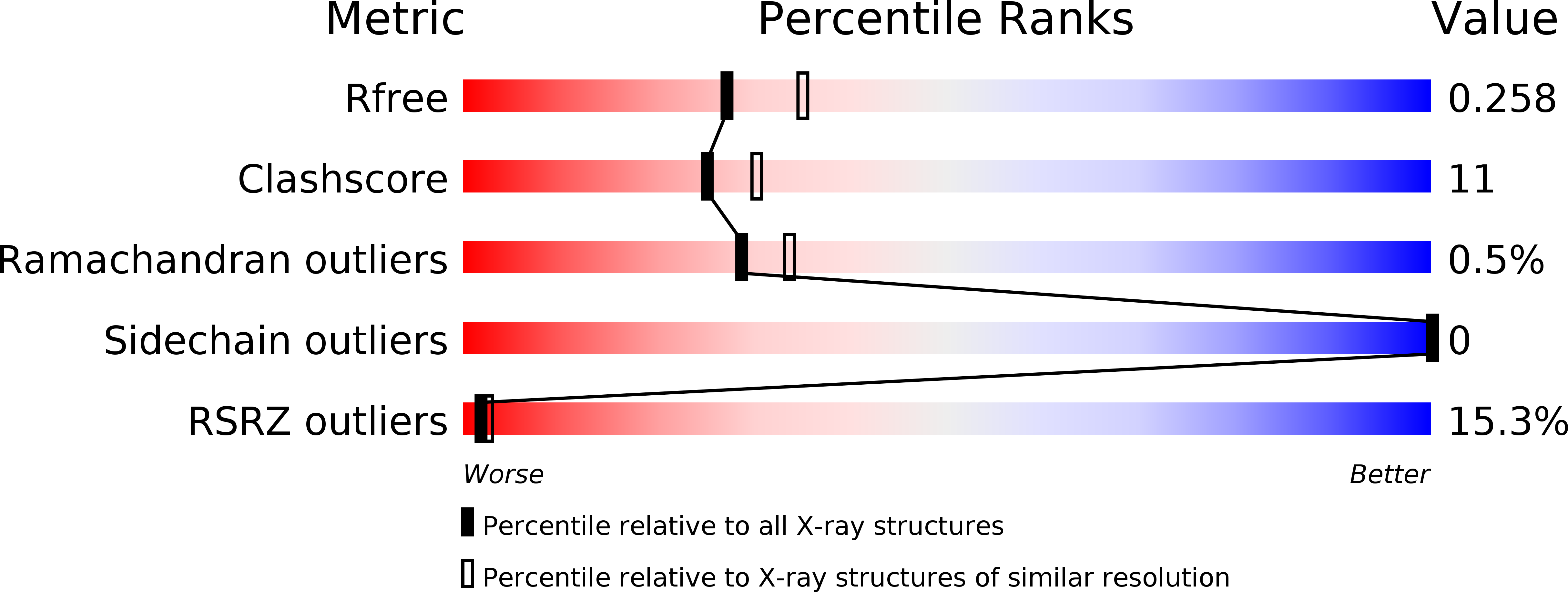
Resolution:
2.28 Å
R-Value Free:
0.26
R-Value Work:
0.22
Space Group:
P 21 21 21