Deposition Date
2013-06-01
Release Date
2016-11-09
Last Version Date
2024-11-06
Entry Detail
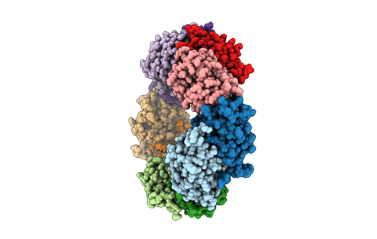
Biological Source:
Source Organism(s):
Plasmodium vivax Sal-1 (Taxon ID: 126793)
Expression System(s):
Method Details:
Experimental Method:
Resolution:
2.50 Å
R-Value Free:
0.21
R-Value Work:
0.17
R-Value Observed:
0.17
Space Group:
P 1 21 1