4L04 image
Entry Detail
PDB ID:
4L04
Keywords:
Title:
Crystal Structure Analysis of human IDH1 mutants in complex with NADP+ and Ca2+/alpha-Ketoglutarate
Biological Source:
Source Organism:
Host Organism:
PDB Version:
Deposition Date:
2013-05-30
Release Date:
2013-07-31
Method Details:
Experimental Method:
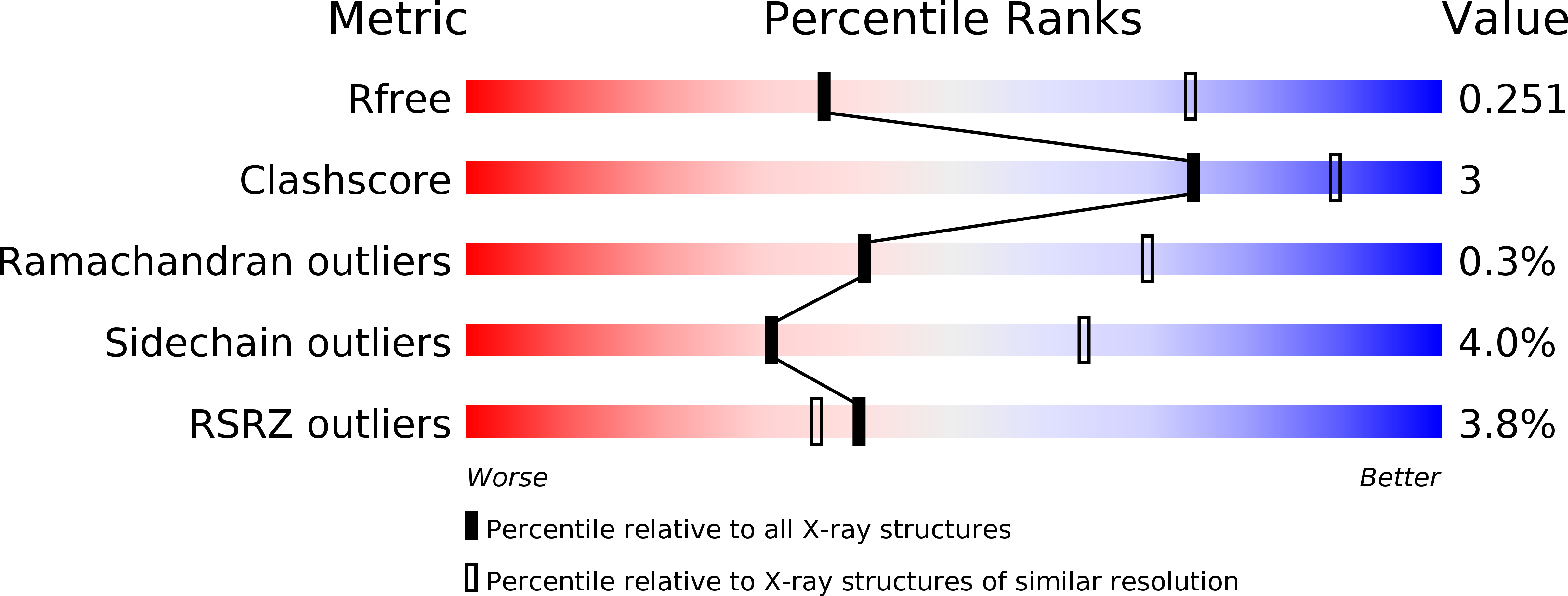
Resolution:
2.87 Å
R-Value Free:
0.25
R-Value Work:
0.19
R-Value Observed:
0.20
Space Group:
P 21 21 21
Macromolecular Entities
Polymer Type:polypeptide(L)
Description:Isocitrate dehydrogenase [NADP] cytoplasmic
Chain IDs:A, B, C, D, E, F
Chain Length:425
Number of Molecules:6
Biological Source:Homo sapiens
Primary Citation
Mutant IDH1 Enhances the Production of 2-Hydroxyglutarate Due to Its Kinetic Mechanism.
Biochemistry 52 4563 4577 (2013)
PMID: 23731180 DOI: 10.1021/bi400514k

Abstact

The human, cytosolic enzyme isocitrate dehydrogenase 1 (IDH1) reversibly converts isocitrate to α-ketoglutarate (αKG). Cancer-associated somatic mutations in IDH1 result in a loss of this normal function but a gain in a new or neomorphic ability to convert αKG to the oncometabolite 2-hydroxyglutarate (2HG). To improve our understanding of the basis for this phenomenon, we have conducted a detailed kinetic study of wild-type IDH1 as well as the known 2HG-producing clinical R132H and G97D mutants and mechanistic Y139D and (newly described) G97N mutants. In the reductive direction of the normal reaction (αKG to isocitrate), dead-end inhibition studies suggest that wild-type IDH1 goes through a random sequential mechanism, similar to previous reports on related mammalian IDH enzymes. However, analogous experiments studying the reductive neomorphic reaction (αKG to 2HG) with the mutant forms of IDH1 are more consistent with an ordered sequential mechanism, with NADPH binding before αKG. This result was further confirmed by primary kinetic isotope effects for which saturating with αKG greatly reduced the observed isotope effect on (D)(V/K)NADPH. For the mutant IDH1 enzyme, the change in mechanism was consistently associated with reduced efficiencies in the use of αKG as a substrate and enhanced efficiencies using NADPH as a substrate. We propose that the sum of these kinetic changes allows the mutant IDH1 enzymes to reductively trap αKG directly into 2HG, rather than allowing it to react with carbon dioxide and form isocitrate, as occurs in the wild-type enzyme.

Legend

Protein

Chemical

Disease

Primary Citation of related structures