
Deposition Date
2013-05-22
Release Date
2013-08-21
Last Version Date
2024-02-28
Entry Detail
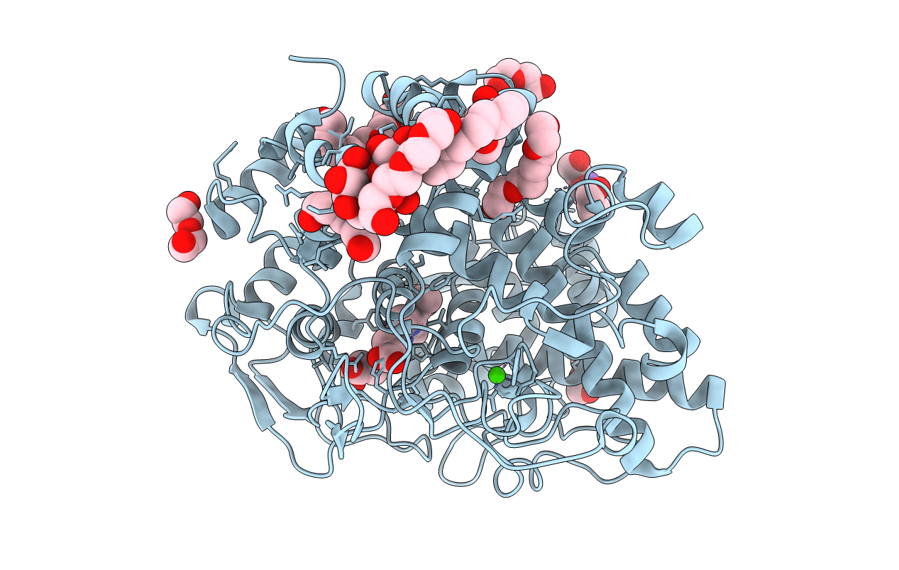
PDB ID:
4KVK
Keywords:
Title:
Crystal structure of Oryza sativa fatty acid alpha-dioxygenase
Biological Source:
Source Organism:
Oryza sativa (Taxon ID: 4530)
Host Organism:
Method Details:
Experimental Method:
Resolution:
1.98 Å
R-Value Free:
0.19
R-Value Work:
0.16
R-Value Observed:
0.16
Space Group:
I 2 2 2
