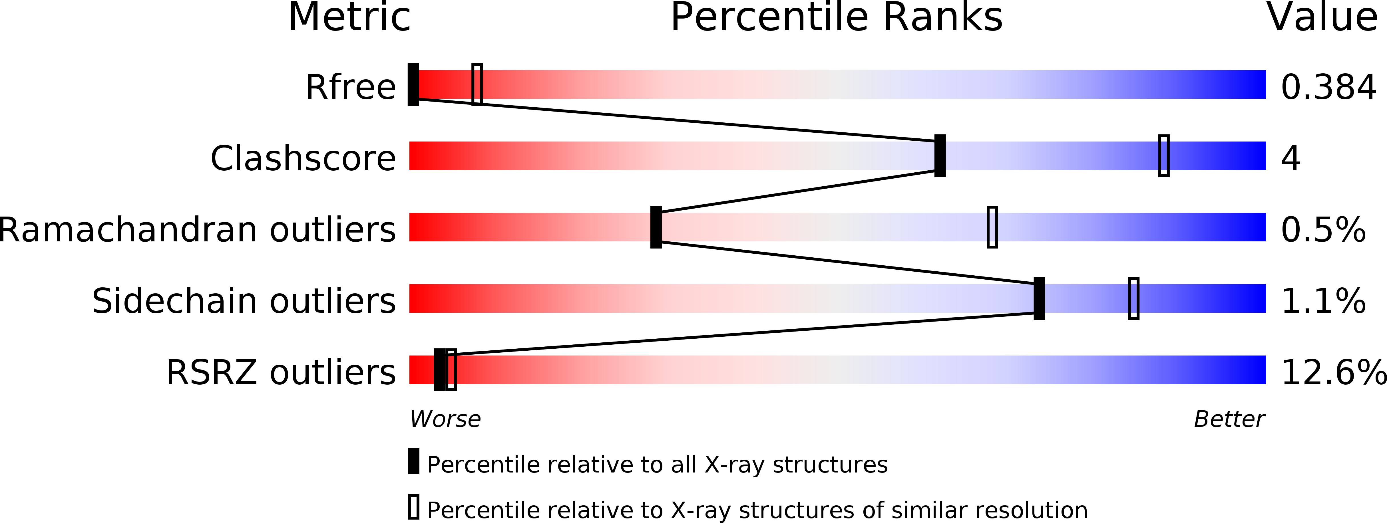
4KSR image
Deposition Date 2013-05-17
Release Date 2013-09-04
Last Version Date 2024-02-28
Entry Detail
PDB ID:
4KSR
Title:
Crystal Structure of the Vibrio cholerae ATPase GspE Hexamer
Biological Source:
Source Organism:
Host Organism:
Method Details:
Experimental Method:
Resolution:
4.20 Å
R-Value Free:
0.37
R-Value Work:
0.38
R-Value Observed:
0.38
Space Group:
P 2 21 21
Macromolecular Entities
Polymer Type:polypeptide(L)
Molecule:Type II secretion system protein E, Hemolysin-coregulated protein
Gene (Uniprot):epsE
Chain IDs:A, B, C
Chain Length:583
Number of Molecules:3
Biological Source:Vibrio cholerae O1, Pseudomonas aeruginosa
Ligand Molecules
Primary Citation
Hexamers of the Type II Secretion ATPase GspE from Vibrio cholerae with Increased ATPase Activity.
Structure 21 1707 1717 (2013)
PMID: 23954505 DOI: 10.1016/j.str.2013.06.027

Abstact

The type II secretion system (T2SS), a multiprotein machinery spanning two membranes in Gram-negative bacteria, is responsible for the secretion of folded proteins from the periplasm across the outer membrane. The critical multidomain T2SS assembly ATPase GspE(EpsE) had not been structurally characterized as a hexamer. Here, four hexamers of Vibrio cholerae GspE(EpsE) are obtained when fused to Hcp1 as an assistant hexamer, as shown with native mass spectrometry. The enzymatic activity of the GspE(EpsE)-Hcp1 fusions is ∼20 times higher than that of a GspE(EpsE) monomer, indicating that increasing the local concentration of GspE(EpsE) by the fusion strategy was successful. Crystal structures of GspE(EpsE)-Hcp1 fusions with different linker lengths reveal regular and elongated hexamers of GspE(EpsE) with major differences in domain orientation within subunits, and in subunit assembly. SAXS studies on GspE(EpsE)-Hcp1 fusions suggest that even further variability in GspE(EpsE) hexamer architecture is likely.

Legend

Protein

Chemical

Disease

Primary Citation of related structures