4KRV image
Deposition Date 2013-05-17
Release Date 2013-08-28
Last Version Date 2024-11-20
Entry Detail
PDB ID:
4KRV
Keywords:
Title:
Crystal structure of catalytic domain of bovine beta1,4-galactosyltransferase mutant M344H-GalT1 complex with 6-sulfo-GlcNAc
Biological Source:
Source Organism:
Bos taurus (Taxon ID: 9913)
Host Organism:
Method Details:
Experimental Method:
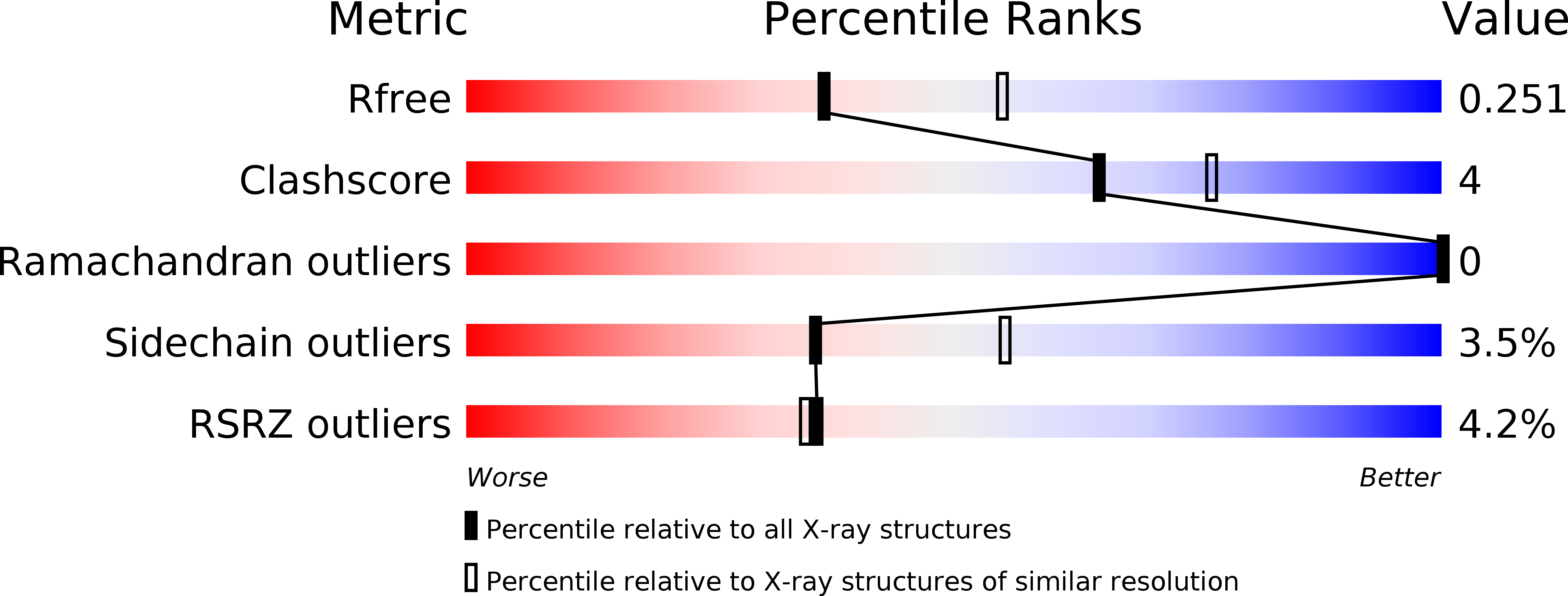
Resolution:
2.40 Å
R-Value Free:
0.24
R-Value Work:
0.18
R-Value Observed:
0.19
Space Group:
P 21 21 21
Macromolecular Entities
Structures with similar UniProt ID
Protein Blast
Polymer Type:polypeptide(L)
Molecule:Beta-1,4-galactosyltransferase 1
Gene (Uniprot):B4GALT1
Mutagens:M344H, C342T
Chain IDs:A, B
Chain Length:286
Number of Molecules:2
Biological Source:Bos taurus
Primary Citation
Investigations on beta 1,4-galactosyltransferase I using 6-sulfo-GlcNAc as an acceptor sugar substrate.
Glycoconj J 30 835 842 (2013)
PMID: 23942731 DOI: 10.1007/s10719-013-9488-4

Abstact

6-sulfate modified N-acetylglucosamine (6-sulfo-GlcNAc) is often found as part of many biologically important carbohydrate epitopes such as 6-sulfo-Le(X). In these epitopes, the 6-sulfo-GlcNAc moiety is extended by a galactose sugar in a β1-4 linkage. The β4GalT1 enzyme transfers galactose (Gal) from UDP-Gal to N-acetylglucosamine (GlcNAc) in the presence of manganese. Here we report that the β4GalT1 enzyme transfers Gal to the 6-sulfo-GlcNAc and 4-methylumbelliferyl-6-sulfo-N-acetyl-β-D-glucosaminide (6-sulfo-βGlcNAc-MU) acceptor substrates, although with very low efficiency. To understand the effect that the 6-sulfate group on the GlcNAc acceptor has on the catalytic activity of the β4GalT1 molecule, we have determined the crystal structure of the catalytic domain of bovine β4GalT1 mutant enzyme M344H-β4GalT1 complex with the 6-sulfo-GlcNAc molecule. In the crystal structure, the 6-sulfo-GlcNAc is bound to the protein in a way that is similar to the GlcNAc molecule. However, the 6-sulfate group engages in additional interactions with the hydrophobic region, residues 276-285, of the protein molecule, and this group is found wedged between the aromatic side chains of Phe-280 and Trp314 residues. Since the side chain of the Trp314 residue undergoes conformational changes during the catalytic cycle of the enzyme, molecular interaction between Trp314 and the 6-sulfate group might hinder this conformational change. Therefore, the lack of a favorable binding environment, together with hindrance to the conformational changes, might be responsible for the poor catalytic activity.

Legend

Protein

Chemical

Disease

Primary Citation of related structures
Feedback Form
Name
Email
Institute
Feedback