4KRR image
Deposition Date 2013-05-16
Release Date 2013-07-10
Last Version Date 2024-10-30
Entry Detail
PDB ID:
4KRR
Title:
Crystal structure of Drosophila WntD N-terminal domain-linker (residues 31-240)
Biological Source:
Source Organism:
Method Details:
Experimental Method:
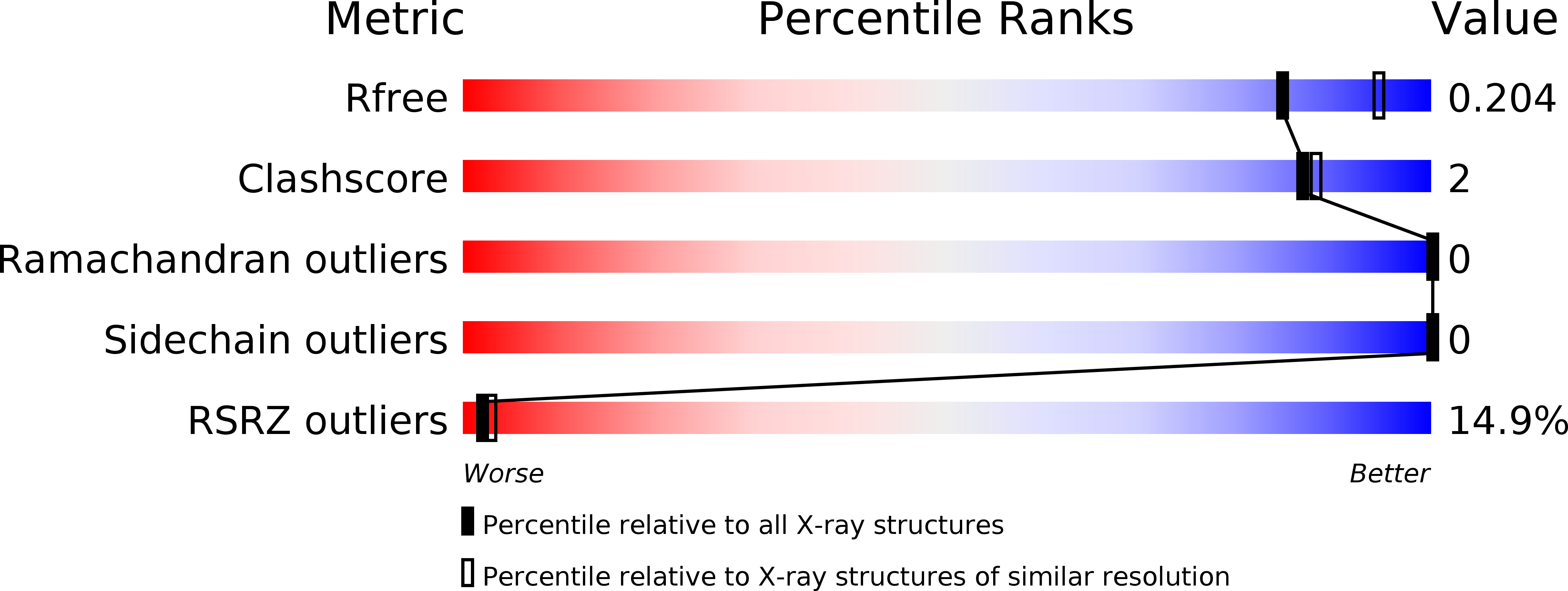
Resolution:
2.12 Å
R-Value Free:
0.20
R-Value Work:
0.17
R-Value Observed:
0.18
Space Group:
P 41
Macromolecular Entities
Structures with similar UniProt ID
Protein Blast
Polymer Type:polypeptide(L)
Molecule:Wnt inhibitor of Dorsal protein
Gene (Uniprot):wntD
Chain IDs:A
Chain Length:221
Number of Molecules:1
Biological Source:Drosophila melanogaster
Primary Citation
Structural Studies of Wnts and Identification of an LRP6 Binding Site.
Structure 21 1235 1242 (2013)
PMID: 23791946 DOI: 10.1016/j.str.2013.05.006

Abstact

Wnts are secreted growth factors that have critical roles in cell fate determination and stem cell renewal. The Wnt/β-catenin pathway is initiated by binding of a Wnt protein to a Frizzled (Fzd) receptor and a coreceptor, LDL receptor-related protein 5 or 6 (LRP5/6). We report the 2.1 Å resolution crystal structure of a Drosophila WntD fragment encompassing the N-terminal domain and the linker that connects it to the C-terminal domain. Differences in the structures of WntD and Xenopus Wnt8, including the positions of a receptor-binding β hairpin and a large solvent-filled cavity in the helical core, indicate conformational plasticity in the N-terminal domain that may be important for Wnt-Frizzled specificity. Structure-based mutational analysis of mouse Wnt3a shows that the linker between the N- and C-terminal domains is required for LRP6 binding. These findings provide important insights into Wnt function and evolution.

Legend

Protein

Chemical

Disease

Primary Citation of related structures