4KQZ image
Deposition Date 2013-05-15
Release Date 2013-07-10
Last Version Date 2024-11-20
Entry Detail
PDB ID:
4KQZ
Keywords:
Title:
structure of the receptor binding domain (RBD) of MERS-CoV spike
Biological Source:
Source Organism:
Host Organism:
Method Details:
Experimental Method:
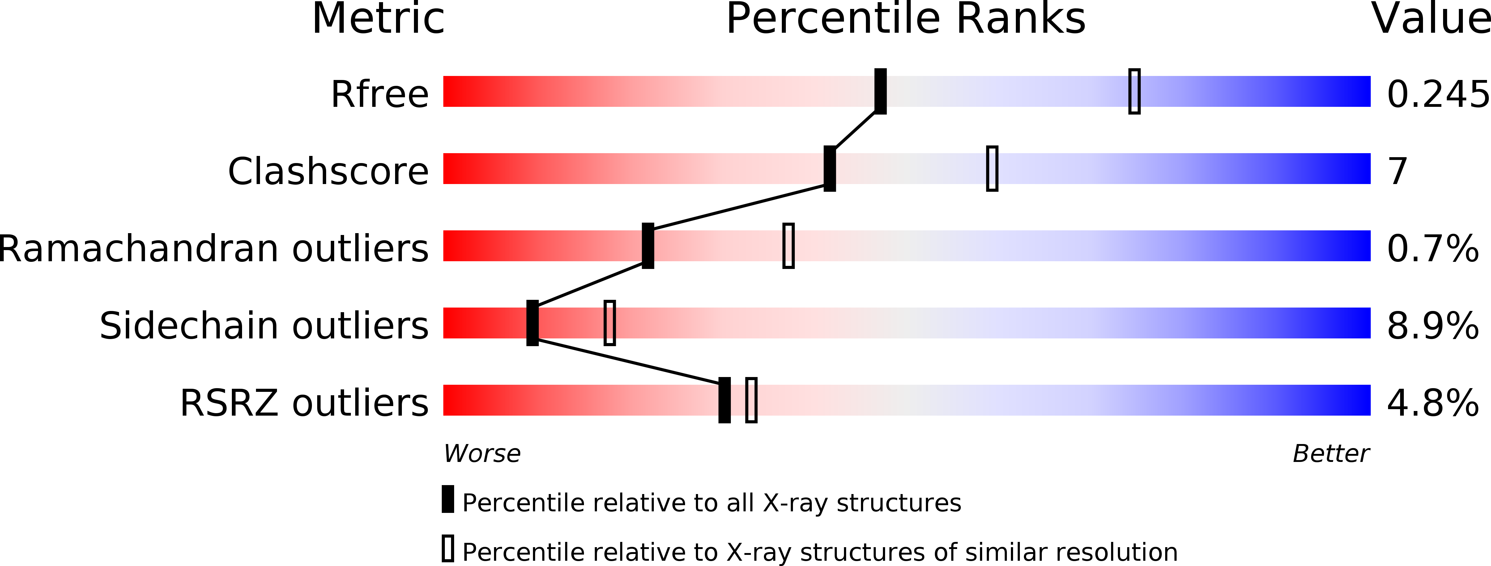
Resolution:
2.51 Å
R-Value Free:
0.25
R-Value Work:
0.20
R-Value Observed:
0.21
Space Group:
P 21 21 21
Macromolecular Entities
Structures with similar UniProt ID
Protein Blast
Polymer Type:polypeptide(L)
Molecule:S protein
Gene (Uniprot):S
Chain IDs:A, B
Chain Length:251
Number of Molecules:2
Biological Source:Human betacoronavirus 2c EMC/2012
Modified Residue
Compound ID Chain ID Parent Comp ID Details 2D Image
ASN A ASN GLYCOSYLATION SITE
Ligand Molecules
Primary Citation
Molecular basis of binding between novel human coronavirus MERS-CoV and its receptor CD26.
Nature 500 227 231 (2013)
PMID: 23831647 DOI: 10.1038/nature12328

Abstact

The newly emergent Middle East respiratory syndrome coronavirus (MERS-CoV) can cause severe pulmonary disease in humans, representing the second example of a highly pathogenic coronavirus, the first being SARS-CoV. CD26 (also known as dipeptidyl peptidase 4, DPP4) was recently identified as the cellular receptor for MERS-CoV. The engagement of the MERS-CoV spike protein with CD26 mediates viral attachment to host cells and virus-cell fusion, thereby initiating infection. Here we delineate the molecular basis of this specific interaction by presenting the first crystal structures of both the free receptor binding domain (RBD) of the MERS-CoV spike protein and its complex with CD26. Furthermore, binding between the RBD and CD26 is measured using real-time surface plasmon resonance with a dissociation constant of 16.7 nM. The viral RBD is composed of a core subdomain homologous to that of the SARS-CoV spike protein, and a unique strand-dominated external receptor binding motif that recognizes blades IV and V of the CD26 β-propeller. The atomic details at the interface between the two binding entities reveal a surprising protein-protein contact mediated mainly by hydrophilic residues. Sequence alignment indicates, among betacoronaviruses, a possible structural conservation for the region homologous to the MERS-CoV RBD core, but a high variation in the external receptor binding motif region for virus-specific pathogenesis such as receptor recognition.

Legend

Protein

Chemical

Disease

Primary Citation of related structures
Feedback Form
Name
Email
Institute
Feedback