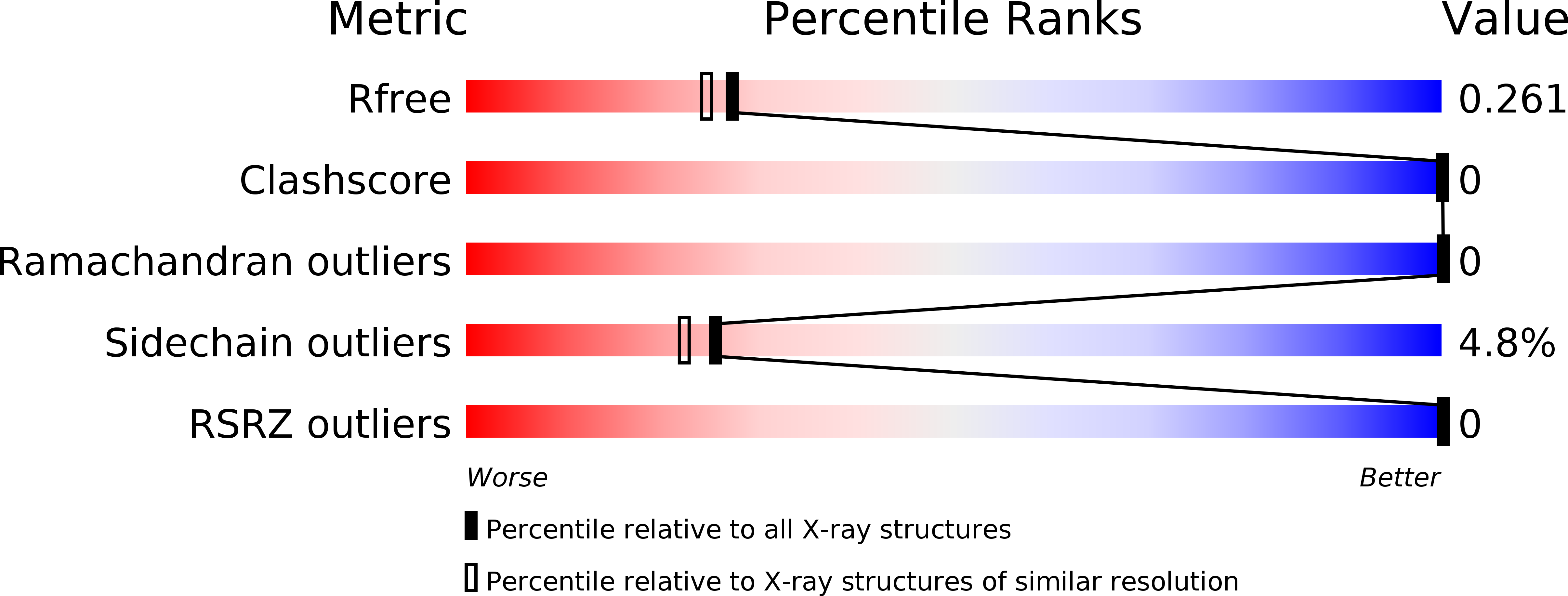
4KMC image
Deposition Date 2013-05-08
Release Date 2014-02-19
Last Version Date 2024-03-20
Entry Detail
PDB ID:
4KMC
Keywords:
Title:
Structure analysis of M. Tuberculosis rRNA transcriptional regulator CarD and its interaction with T. Aquaticus RNA polymerase-BETA1 domain
Biological Source:
Source Organism:
Host Organism:
Method Details:
Experimental Method:
Resolution:
2.15 Å
R-Value Free:
0.25
R-Value Work:
0.21
R-Value Observed:
0.22
Space Group:
P 43 21 2
Macromolecular Entities
Structures with similar UniProt ID
Protein Blast
Polymer Type:polypeptide(L)
Molecule:RNA polymerase-binding transcription factor CarD
Chain IDs:A
Chain Length:80
Number of Molecules:1
Biological Source:Mycobacterium tuberculosis
Primary Citation
Structure of the carboxy-terminal domain of Mycobacterium tuberculosis CarD protein: an essential rRNA transcriptional regulator.
Acta Crystallogr.,Sect.F 70 160 165 (2014)
PMID: 24637748 DOI: 10.1107/S2053230X13034407

Abstact

The CarD protein is highly expressed in mycobacterial strains under basal conditions and is transcriptionally induced during multiple types of genotoxic stress and starvation. The CarD protein binds the β subunit of RNA polymerase and influences gene expression. The disruption of interactions between CarD and the β subunit of RNA polymerase has a significant effect on mycobacterial survival, resistance to stress and pathogenesis. To understand the structure of CarD and its interaction with the β subunit of RNA polymerase, Mycobacterium tuberculosis CarD (MtbCarD) and the Thermus aquaticus RNA polymerase β subunit were recombinantly expressed and purified. Secondary-structure analysis using circular-dichroism spectroscopy indicated that MtbCarD contains ∼ 60% α-helix, ∼ 7% β-sheet and ∼ 33% random-coil structure. The C-terminal domain of MtbCarD (CarD(83-161)) was crystallized and its X-ray structure was determined at 2.1 Å resolution. CarD(83-161) forms a distorted Y-shaped structure containing bundles of three helices connected by a loop. The residues forming the distorted Y-shaped structure are highly conserved in CarD sequences from other mycobacterial species. Comparison of the CarD(83-161) structure with the recently determined full-length M. tuberculosis and T. thermophilus CarD crystal structures revealed structural differences in residues 141-161 of the C-terminal domain of the CarD(83-161) structure. The structural changes in the CarD(83-161) structure occurred owing to proteolysis and crystallization artifacts.

Legend

Protein

Chemical

Disease

Primary Citation of related structures