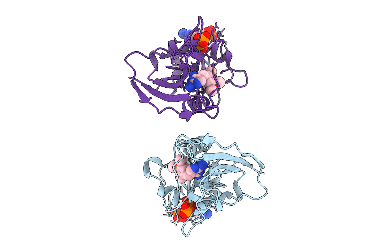
4KM2 image
Entry Detail
PDB ID:
4KM2
Keywords:
Title:
Crystal structure of Dihydrofolate reductase from Mycobacterium tuberculosis in an open conformation in complex with trimethoprim
Biological Source:
Host Organism:
PDB Version:
Deposition Date:
2013-05-07
Release Date:
2013-12-25
Method Details:
Experimental Method:
Resolution:
1.40 Å
R-Value Free:
0.20
R-Value Work:
0.14
R-Value Observed:
0.14
Space Group:
P 21 21 21
Macromolecular Entities
Polymer Type:polypeptide(L)
Description:Dihydrofolate reductase
Chain IDs:A, B
Chain Length:179
Number of Molecules:2
Biological Source:Mycobacterium tuberculosis
Primary Citation
Mycobacterium tuberculosis Dihydrofolate Reductase Reveals Two Conformational States and a Possible Low Affinity Mechanism to Antifolate Drugs.
Structure 22 94 103 (2014)
PMID: 24210757 DOI: 10.1016/j.str.2013.09.022

Abstact

Inhibition of the biosynthesis of tetrahydrofolate (THF) has long been a focus in the treatment of both cancer and infectious diseases. Dihydrofolate reductase (DHFR), which catalyzes the last step, is one of the most thoroughly explored targets of this pathway, but there are no DHFR inhibitors used for tuberculosis treatment. Here, we report a structural, site-directed mutagenesis and calorimetric analysis of Mycobacterium tuberculosis DHFR (MtDHFR) in complex with classical DHFR inhibitors. Our study provides insights into the weak inhibition of MtDHFR by trimethoprim and other antifolate drugs, such as pyrimethamine and cycloguanil. The construction of the mutant Y100F, together with calorimetric studies, gives insights into low affinity of MtDHFR for classical DHFR inhibitors. Finally, the structures of MtDHFR in complex with pyrimethamine and cycloguanil define important interactions in the active site and provide clues to the more effective design of antibiotics targeted against MtDHFR.

Legend

Protein

Chemical

Disease

Primary Citation of related structures