
Deposition Date
2013-04-29
Release Date
2013-05-08
Last Version Date
2023-12-06
Entry Detail
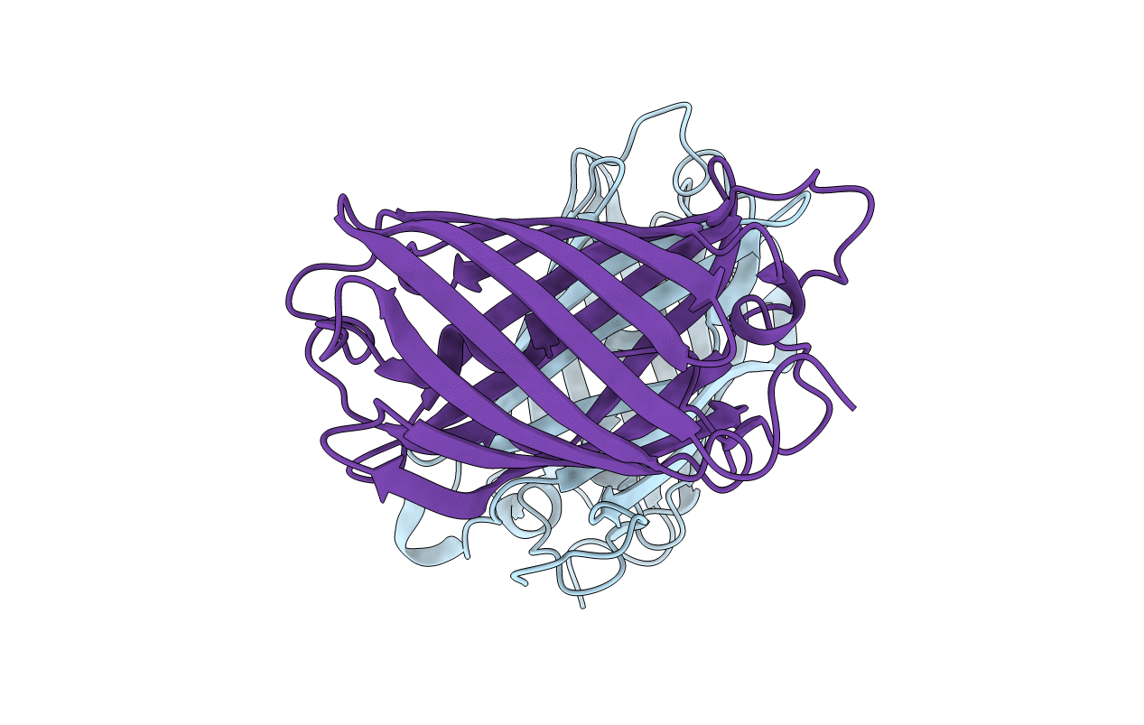
PDB ID:
4KGE
Keywords:
Title:
Crystal structure of near-infrared fluorescent protein with an extended stokes shift, pH 4.5
Biological Source:
Source Organism(s):
synthetic construct (Taxon ID: 32630)
Expression System(s):
Method Details:
Experimental Method:
Resolution:
2.30 Å
R-Value Free:
0.23
R-Value Work:
0.18
R-Value Observed:
0.18
Space Group:
P 61 2 2
