4KGB image
Deposition Date 2013-04-29
Release Date 2013-10-23
Last Version Date 2023-11-08
Entry Detail
PDB ID:
4KGB
Keywords:
Title:
Structure of succinyl-CoA: 3-ketoacid CoA transferase from Drosophila melanogaster
Biological Source:
Source Organism:
Host Organism:
Method Details:
Experimental Method:
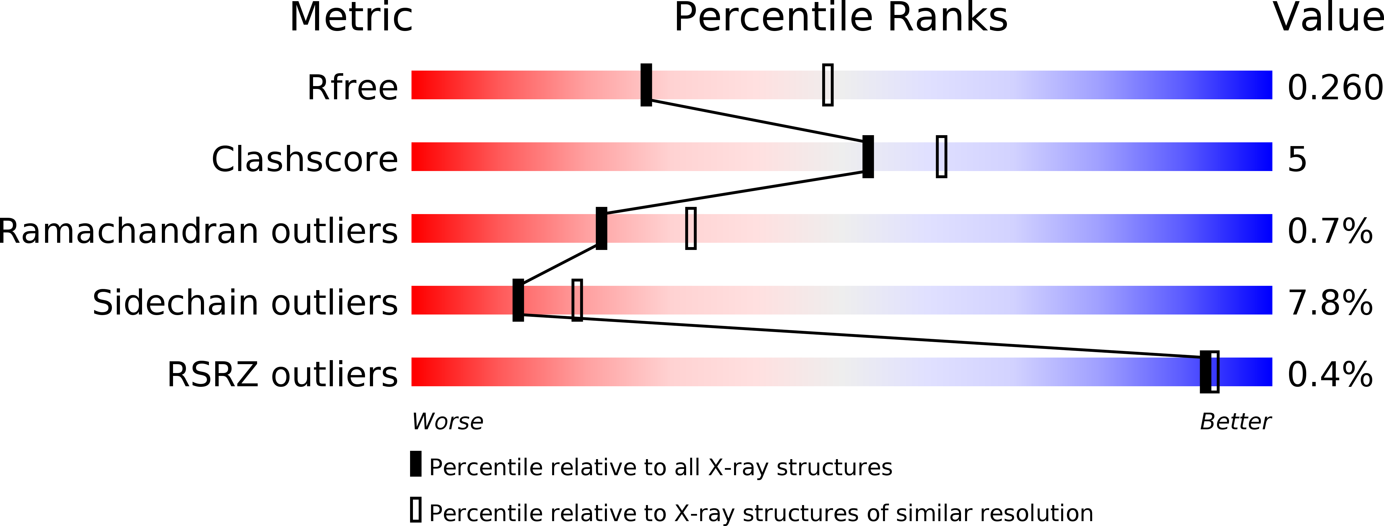
Resolution:
2.64 Å
R-Value Free:
0.26
R-Value Work:
0.20
R-Value Observed:
0.20
Space Group:
P 21 21 21
Macromolecular Entities
Structures with similar UniProt ID
Protein Blast
Polymer Type:polypeptide(L)
Molecule:Succinyl-CoA:3-ketoacid-coenzyme A transferase
Gene (Uniprot):Scot
Chain IDs:A, B
Chain Length:487
Number of Molecules:2
Biological Source:Drosophila melanogaster
Ligand Molecules
Primary Citation
Structure of succinyl-CoA:3-ketoacid CoA transferase from Drosophila melanogaster.
Acta Crystallogr.,Sect.F 69 1089 1093 (2013)
PMID: 24100554 DOI: 10.1107/S1744309113024986

Abstact

Succinyl-CoA:3-ketoacid CoA transferase (SCOT) plays a crucial role in ketone-body metabolism. SCOT from Drosophila melanogaster (DmSCOT) was purified and crystallized. The crystal structure of DmSCOT was determined at 2.64 Å resolution and belonged to space group P212121, with unit-cell parameters a=76.638, b=101.921, c=122.457 Å, α=β=γ=90°. Sequence alignment and structural analysis identified DmSCOT as a class I CoA transferase. Compared with Acetobacter aceti succinyl-CoA:acetate CoA transferase, DmSCOT has a different substrate-binding pocket, which may explain the difference in their substrate specificities.

Legend

Protein

Chemical

Disease

Primary Citation of related structures