
Deposition Date
2013-04-24
Release Date
2013-07-31
Last Version Date
2024-10-16
Entry Detail
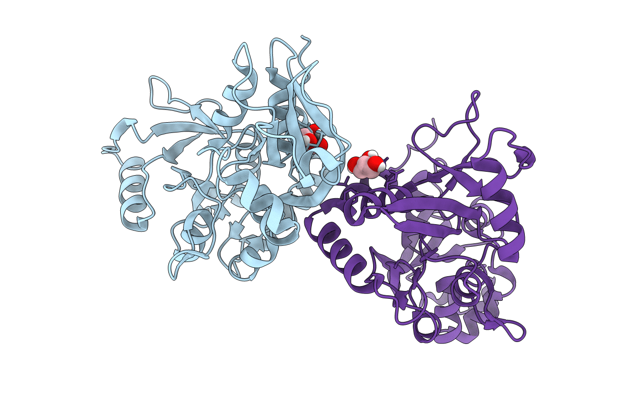
PDB ID:
4KCD
Keywords:
Title:
Crystal Structure of the NMDA Receptor GluN3A Ligand Binding Domain Apo State
Biological Source:
Source Organism(s):
Rattus norvegicus (Taxon ID: 10116)
Expression System(s):
Method Details:
Experimental Method:
Resolution:
1.68 Å
R-Value Free:
0.18
R-Value Work:
0.15
R-Value Observed:
0.15
Space Group:
P 21 21 21
