4KBM image
Entry Detail
PDB ID:
4KBM
Title:
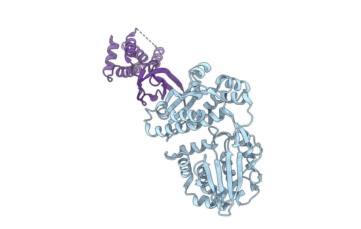
Structure of the Mtb CarD/RNAP Beta subunit B1-B2 domains complex
Biological Source:
Host Organism:
PDB Version:
Deposition Date:
2013-04-23
Release Date:
2013-10-02
Method Details:
Experimental Method:
Resolution:
2.11 Å
R-Value Free:
0.23
R-Value Work:
0.20
R-Value Observed:
0.20
Space Group:
C 2 2 21
Macromolecular Entities
Polymer Type:polypeptide(L)
Description:DNA-directed RNA polymerase subunit beta
Chain IDs:A
Chain Length:409
Number of Molecules:1
Biological Source:Mycobacterium tuberculosis
Polymer Type:polypeptide(L)
Description:RNA polymerase-binding transcription factor CarD
Chain IDs:B
Chain Length:162
Number of Molecules:1
Biological Source:Mycobacterium tuberculosis
Primary Citation
Structure of the Mtb CarD/RNAP beta-Lobes Complex Reveals the Molecular Basis of Interaction and Presents a Distinct DNA-Binding Domain for Mtb CarD.
Structure 21 1859 1869 (2013)
PMID: 24055315 DOI: 10.1016/j.str.2013.08.014

Abstact

CarD from Mycobacterium tuberculosis (Mtb) is an essential protein shown to be involved in stringent response through downregulation of rRNA and ribosomal protein genes. CarD interacts with the β-subunit of RNAP and this interaction is vital for Mtb's survival during the persistent infection state. We have determined the crystal structure of CarD in complex with the RNAP β-subunit β1 and β2 domains at 2.1 Å resolution. The structure reveals the molecular basis of CarD/RNAP interaction, providing a basis to further our understanding of RNAP regulation by CarD. The structural fold of the CarD N-terminal domain is conserved in RNAP interacting proteins such as TRCF-RID and CdnL, and displays similar interactions to the predicted homology model based on the TRCF/RNAP β1 structure. Interestingly, the structure of the C-terminal domain, which is required for complete CarD function in vivo, represents a distinct DNA-binding fold.

Legend

Protein

Chemical

Disease

Primary Citation of related structures