4K9J image
Entry Detail
PDB ID:
4K9J
Title:
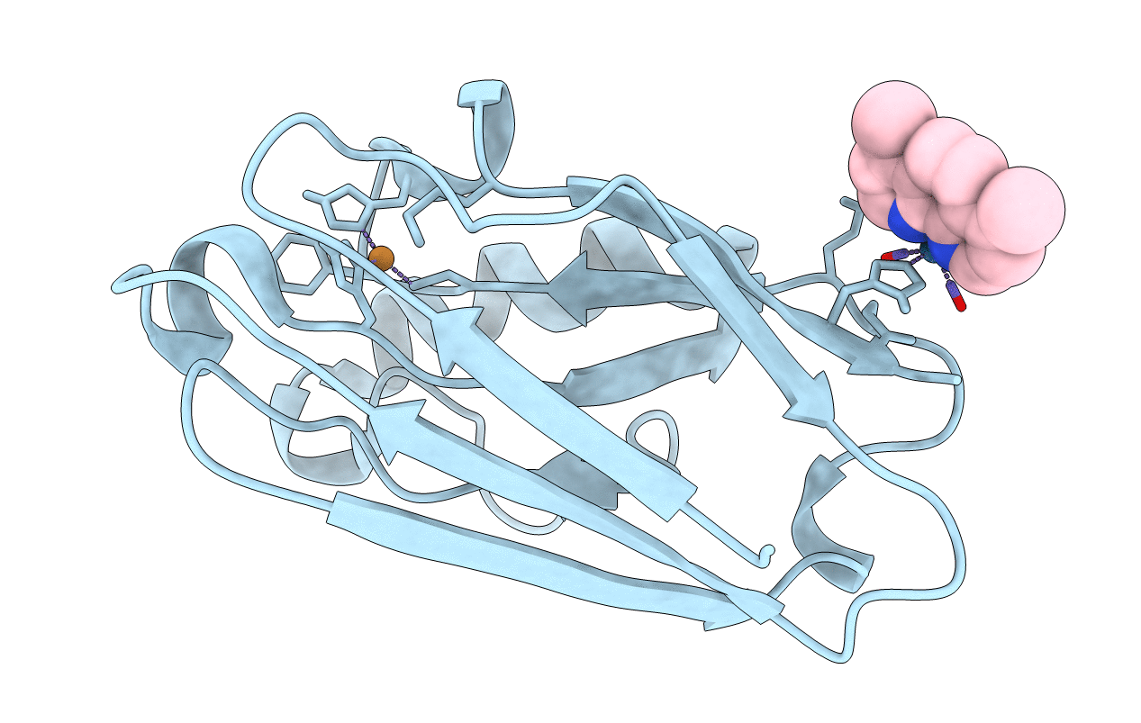
Structure of Re(CO)3(4,7-dimethyl-phen)(Thr126His)(Lys122Trp)(His83Glu)(Trp48Phe)(Tyr72Phe)(Tyr108Phe)AzCu(II), a Rhenium modified Azurin mutant
Biological Source:
Source Organism:
Host Organism:
PDB Version:
Deposition Date:
2013-04-20
Release Date:
2013-10-02
Method Details:
Experimental Method:
Resolution:
1.70 Å
R-Value Free:
0.23
R-Value Work:
0.19
R-Value Observed:
0.19
Space Group:
F 2 2 2
Macromolecular Entities
Polymer Type:polypeptide(L)
Description:Azurin
Mutations:t126H, k122w, h83e,w48f, y72f
Chain IDs:A
Chain Length:128
Number of Molecules:1
Biological Source:Pseudomonas aeruginosa
Primary Citation
Tryptophan-accelerated electron flow across a protein-protein interface.
J.Am.Chem.Soc. 135 15515 15525 (2013)
PMID: 24032375 DOI: 10.1021/ja406830d

Abstact

We report a new metallolabeled blue copper protein, Re126W122Cu(I) Pseudomonas aeruginosa azurin, which has three redox sites at well-defined distances in the protein fold: Re(I)(CO)3(4,7-dimethyl-1,10-phenanthroline) covalently bound at H126, a Cu center, and an indole side chain W122 situated between the Re and Cu sites (Re-W122(indole) = 13.1 Å, dmp-W122(indole) = 10.0 Å, Re-Cu = 25.6 Å). Near-UV excitation of the Re chromophore leads to prompt Cu(I) oxidation (<50 ns), followed by slow back ET to regenerate Cu(I) and ground-state Re(I) with biexponential kinetics, 220 ns and 6 μs. From spectroscopic measurements of kinetics and relative ET yields at different concentrations, it is likely that the photoinduced ET reactions occur in protein dimers, (Re126W122Cu(I))2 and that the forward ET is accelerated by intermolecular electron hopping through the interfacial tryptophan: *Re//←W122←Cu(I), where // denotes a protein-protein interface. Solution mass spectrometry confirms a broad oligomer distribution with prevalent monomers and dimers, and the crystal structure of the Cu(II) form shows two Re126W122Cu(II) molecules oriented such that redox cofactors Re(dmp) and W122-indole on different protein molecules are located at the interface at much shorter intermolecular distances (Re-W122(indole) = 6.9 Å, dmp-W122(indole) = 3.5 Å, and Re-Cu = 14.0 Å) than within single protein folds. Whereas forward ET is accelerated by hopping through W122, BET is retarded by a space jump at the interface that lacks specific interactions or water molecules. These findings on interfacial electron hopping in (Re126W122Cu(I))2 shed new light on optimal redox-unit placements required for functional long-range charge separation in protein complexes.

Legend

Protein

Chemical

Disease

Primary Citation of related structures