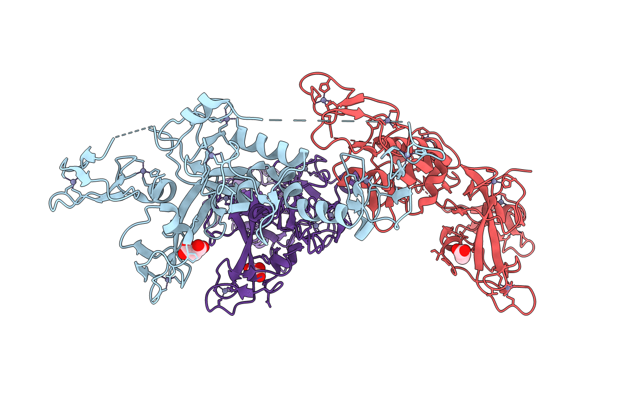
4K7D image
Deposition Date 2013-04-17
Release Date 2013-05-15
Last Version Date 2024-02-28
Entry Detail
PDB ID:
4K7D
Keywords:
Title:
Crystal Structure of Parkin C-terminal RING domains
Biological Source:
Source Organism:
Host Organism:
Method Details:
Experimental Method:
Resolution:
2.80 Å
R-Value Free:
0.23
R-Value Work:
0.18
R-Value Observed:
0.18
Space Group:
P 21 21 21
Macromolecular Entities
Polymer Type:polypeptide(L)
Molecule:E3 ubiquitin-protein ligase parkin
Gene (Uniprot):Prkn
Chain IDs:A, B, C
Chain Length:330
Number of Molecules:3
Biological Source:Rattus norvegicus
Primary Citation
Structure of parkin reveals mechanisms for ubiquitin ligase activation.
Science 340 1451 1455 (2013)
PMID: 23661642 DOI: 10.1126/science.1237908

Abstact

Mutations in the PARK2 (parkin) gene are responsible for an autosomal recessive form of Parkinson's disease. The parkin protein is a RING-in-between-RING E3 ubiquitin ligase that exhibits low basal activity. We describe the crystal structure of full-length rat parkin. The structure shows parkin in an autoinhibited state and provides insight into how it is activated. RING0 occludes the ubiquitin acceptor site Cys(431) in RING2, whereas a repressor element of parkin binds RING1 and blocks its E2-binding site. Mutations that disrupted these inhibitory interactions activated parkin both in vitro and in cells. Parkin is neuroprotective, and these findings may provide a structural and mechanistic framework for enhancing parkin activity.

Legend

Protein

Chemical

Disease

Primary Citation of related structures