4K2E image
Deposition Date 2013-04-09
Release Date 2014-04-30
Last Version Date 2024-11-13
Entry Detail
PDB ID:
4K2E
Title:
HlyU from Vibrio cholerae N16961
Biological Source:
Source Organism:
Host Organism:
Method Details:
Experimental Method:
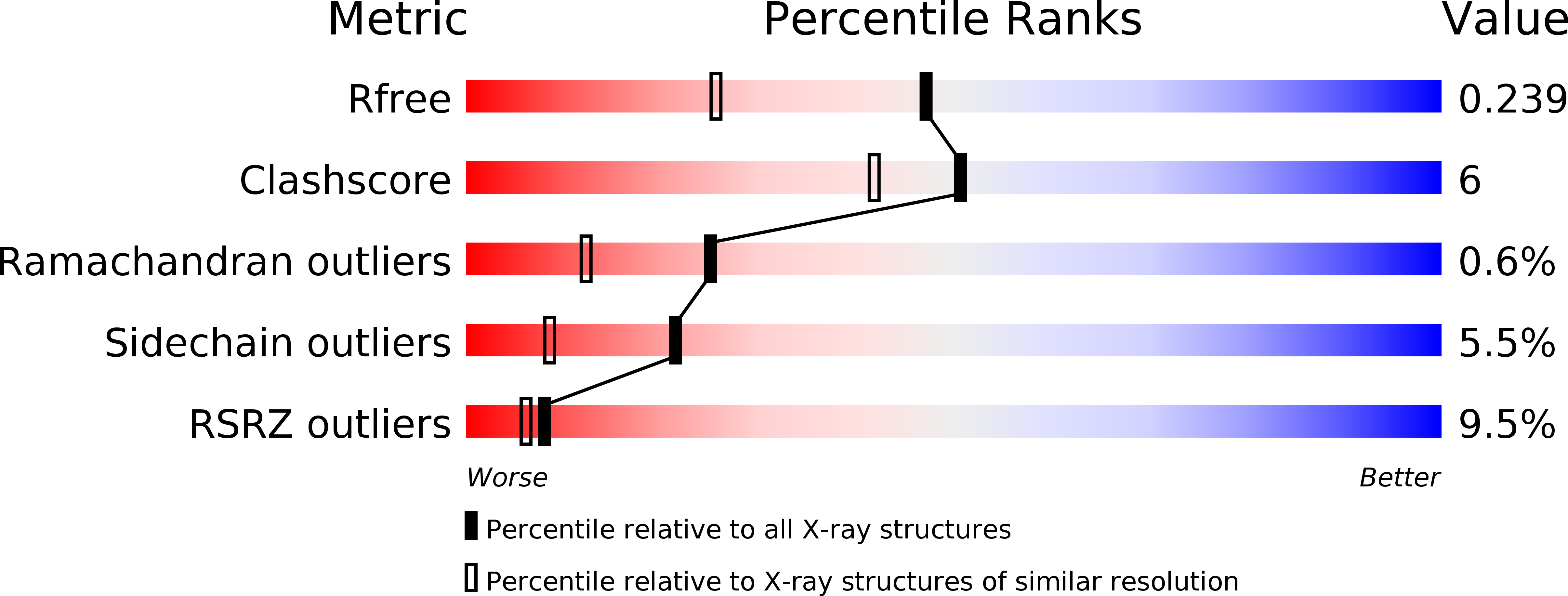
Resolution:
1.80 Å
R-Value Free:
0.23
R-Value Work:
0.18
R-Value Observed:
0.19
Space Group:
C 1 2 1
Macromolecular Entities
Structures with similar UniProt ID
Protein Blast
Polymer Type:polypeptide(L)
Molecule:Transcriptional activator HlyU
Gene (Uniprot):hlyU
Chain IDs:A, B, C, D
Chain Length:112
Number of Molecules:4
Biological Source:Vibrio cholerae
Modified Residue
Compound ID Chain ID Parent Comp ID Details 2D Image
CSO A CYS S-HYDROXYCYSTEINE
Primary Citation
Crystal structure of HlyU, the hemolysin gene transcription activator, from Vibrio cholerae N16961 and functional implications.
Biochim.Biophys.Acta 1844 2346 2354 (2014)
PMID: 25450504 DOI: 10.1016/j.bbapap.2014.09.020

Abstact

HlyU in Vibrio cholerae is known to be the transcriptional activator of the hemolysin gene, HlyA and possibly a regulator of other virulence factors influencing growth, colonization and pathogenicity of this infective agent. Here we report the crystal structure of HlyU from V. cholerae N16961 (HlyU_Vc) at 1.8Å. The protein, with five α-helices and three β-strands in the topology of α1-α2-β1-α3-α4-β2-β3-α5, forms a homodimer. Helices α3-α4 and a β sheet form the winged helix-turn-helix (wHTH) DNA-binding motif common to the transcription regulators of the SmtB/ArsR family. In spite of an overall fold similar to SmtB/ArsR family, it lacks any metal binding site seen in SmtB. A comparison of the dimeric interfaces showed that the one in SmtB is much larger and have salt bridges that can be disrupted to accommodate metal ions. A model of HlyU-DNA complex suggests bending of the DNA. Cys38 in the structure was found to be modified as sulfenic acid; the oxidized form was not seen in another structure solved under reducing condition. Although devoid of any metal binding site, the presence of a Cys residue exhibiting oxidation-reduction suggests the possibility of the existence of a redox switch in transcription regulation. A structure-based phylogenetic analysis of wHTH proteins revealed the segregation of metal and non-metal binding proteins as well as those in the latter group that are under redox control.

Legend

Protein

Chemical

Disease

Primary Citation of related structures
Feedback Form
Name
Email
Institute
Feedback