4K28 image
Deposition Date 2013-04-08
Release Date 2013-04-17
Last Version Date 2024-02-28
Entry Detail
PDB ID:
4K28
Keywords:
Title:
2.15 Angstrom resolution crystal structure of a shikimate dehydrogenase family protein from Pseudomonas putida KT2440 in complex with NAD+
Biological Source:
Source Organism:
Host Organism:
Method Details:
Experimental Method:
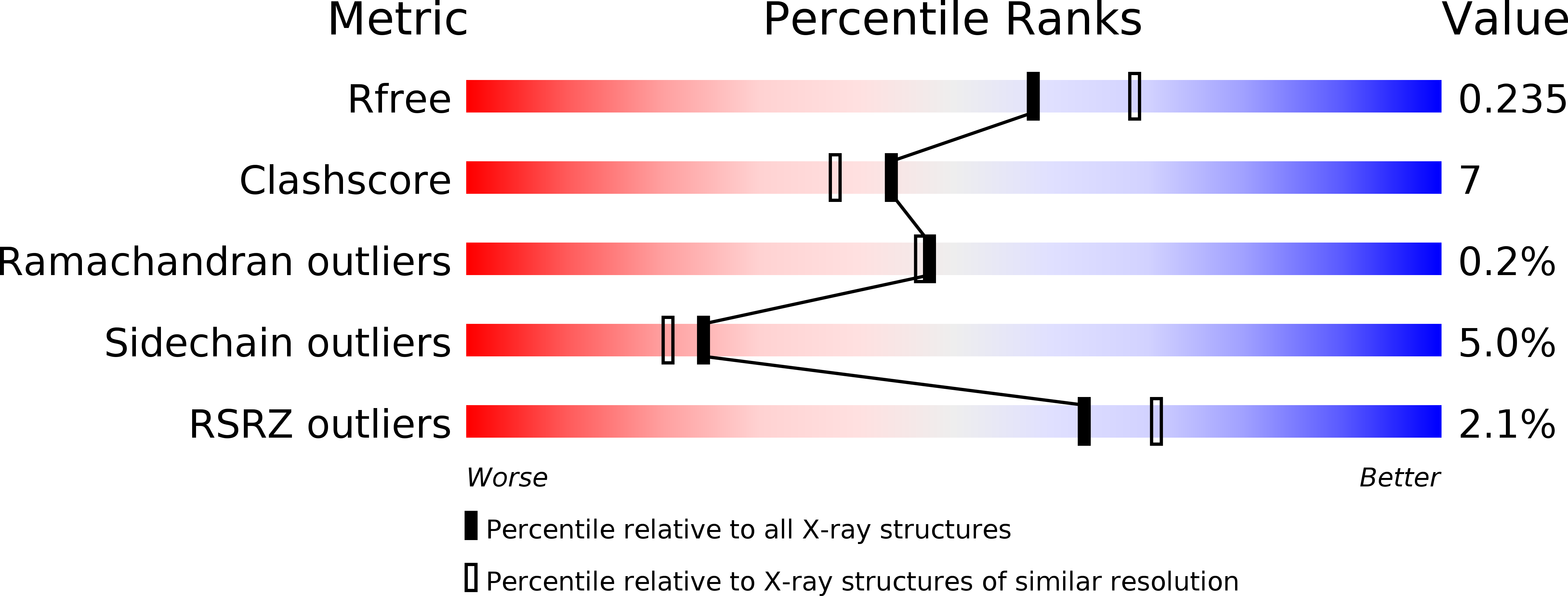
Resolution:
2.15 Å
R-Value Free:
0.23
R-Value Work:
0.17
R-Value Observed:
0.18
Space Group:
P 21 21 21
Macromolecular Entities
Structures with similar UniProt ID
Protein Blast
Polymer Type:polypeptide(L)
Molecule:Shikimate dehydrogenase family protein
Gene (Uniprot):PP_2608
Chain IDs:A, B
Chain Length:269
Number of Molecules:2
Biological Source:Pseudomonas putida
Primary Citation
Insights into the function of RifI2: structural and biochemical investigation of a new shikimate dehydrogenase family protein.
Biochim.Biophys.Acta 1834 516 523 (2013)
PMID: 23142411 DOI: 10.1016/j.bbapap.2012.10.016

Abstact

The shikimate dehydrogenase (SDH) family consists of enzymes with diverse roles in secondary metabolism. The two most widespread members of the family, AroE and YdiB, function in amino acid biosynthesis and quinate catabolism, respectively. Here, we have determined the crystal structure of an SDH homolog belonging to the RifI class, a group of enzymes with proposed roles in antibiotic biosynthesis. The structure of RifI2 from Pseudomonas putida exhibits a number of distinctive features, including a substantial C-terminal truncation and an atypical mode of oligomerization. The active site of the enzyme contains substrate- and cofactor-binding motifs that are significantly different from those of any previously characterized member of the SDH family. These features are reflected in the novel kinetic properties of the enzyme. RifI2 exhibits much lower activity using shikimate as a substrate than AroE, and a strong preference for NAD(+) instead of NADP(+) as a cofactor. Moreover, the enzyme has only trace activity using quinate, unlike YdiB. Cocrystallization of RifI2 with NAD(+) provided the opportunity to determine the mode of cofactor selectivity employed by the enzyme. We complemented this analysis by probing the role of a strictly conserved residue in the cofactor-binding domain, Asn193, by site directed mutagenesis. This study presents the first crystal structure and formal kinetic characterization of a new NAD(+)-dependent member of the SDH family.

Legend

Protein

Chemical

Disease

Primary Citation of related structures
Feedback Form
Name
Email
Institute
Feedback