4K0K image
Deposition Date 2013-04-04
Release Date 2013-06-26
Last Version Date 2024-10-09
Entry Detail
PDB ID:
4K0K
Keywords:
Title:
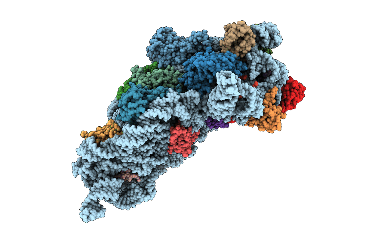
Crystal structure of the Thermus thermophilus 30S ribosomal subunit complexed with a serine-ASL and mRNA containing a stop codon
Biological Source:
Source Organism:
Method Details:
Experimental Method:
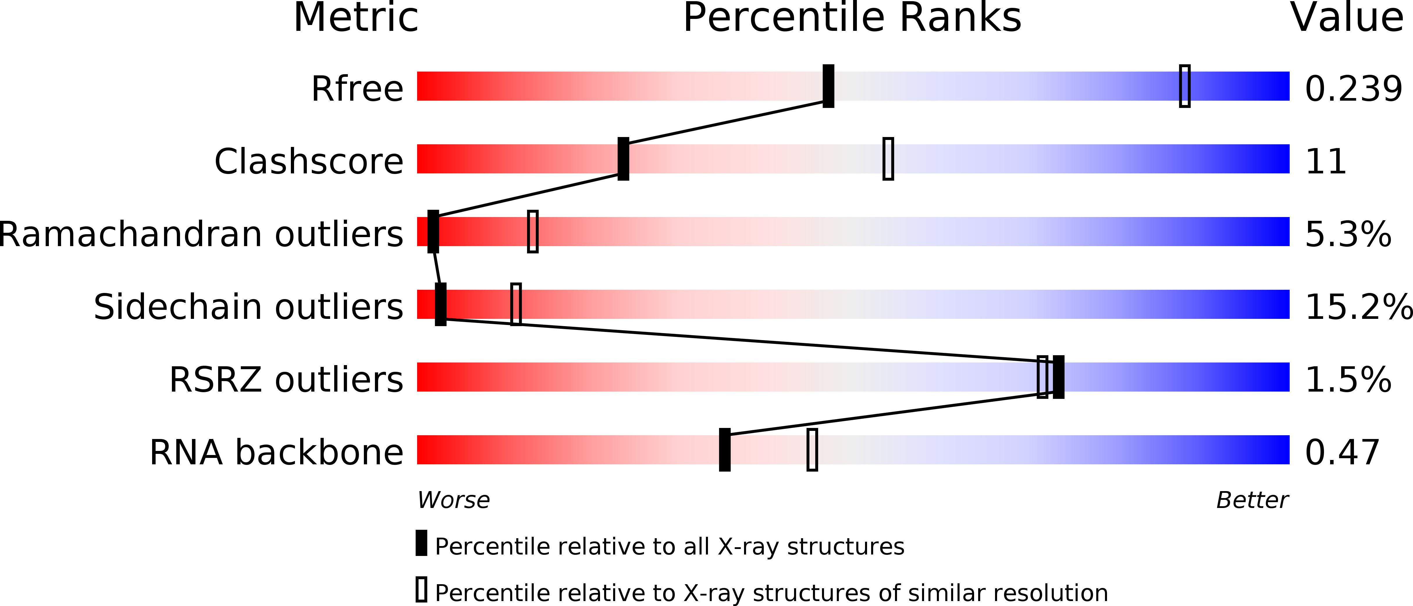
Resolution:
3.40 Å
R-Value Free:
0.24
R-Value Work:
0.18
R-Value Observed:
0.19
Space Group:
P 41 21 2
Macromolecular Entities
Polymer Type:polyribonucleotide
Molecule:16S ribosomal RNA
Chain IDs:A
Chain Length:1517
Number of Molecules:1
Biological Source:Thermus thermophilus
Structures with similar UniProt ID
Protein Blast
Polymer Type:polypeptide(L)
Molecule:30S ribosomal protein S2
Gene (Uniprot):rpsB
Chain IDs:B
Chain Length:235
Number of Molecules:1
Biological Source:Thermus thermophilus
Structures with similar UniProt ID
Protein Blast
Polymer Type:polypeptide(L)
Molecule:30S ribosomal protein S3
Gene (Uniprot):rpsC
Chain IDs:C
Chain Length:207
Number of Molecules:1
Biological Source:Thermus thermophilus
Structures with similar UniProt ID
Protein Blast
Polymer Type:polypeptide(L)
Molecule:30S ribosomal protein S4
Gene (Uniprot):rpsD
Chain IDs:D
Chain Length:208
Number of Molecules:1
Biological Source:Thermus thermophilus
Structures with similar UniProt ID
Protein Blast
Polymer Type:polypeptide(L)
Molecule:30S ribosomal protein S5
Gene (Uniprot):rpsE
Chain IDs:E
Chain Length:151
Number of Molecules:1
Biological Source:Thermus thermophilus
Structures with similar UniProt ID
Protein Blast
Polymer Type:polypeptide(L)
Molecule:30S ribosomal protein S6
Gene (Uniprot):rpsF
Chain IDs:F
Chain Length:101
Number of Molecules:1
Biological Source:Thermus thermophilus
Structures with similar UniProt ID
Protein Blast
Polymer Type:polypeptide(L)
Molecule:30S ribosomal protein S7
Gene (Uniprot):rpsG
Chain IDs:G
Chain Length:155
Number of Molecules:1
Biological Source:Thermus thermophilus
Structures with similar UniProt ID
Protein Blast
Polymer Type:polypeptide(L)
Molecule:30S ribosomal protein S8
Chain IDs:H
Chain Length:138
Number of Molecules:1
Biological Source:Thermus thermophilus
Structures with similar UniProt ID
Protein Blast
Polymer Type:polypeptide(L)
Molecule:30S ribosomal protein S9
Gene (Uniprot):rpsI
Chain IDs:I
Chain Length:127
Number of Molecules:1
Biological Source:Thermus thermophilus
Structures with similar UniProt ID
Protein Blast
Polymer Type:polypeptide(L)
Molecule:30S ribosomal protein S10
Gene (Uniprot):rpsJ
Chain IDs:J
Chain Length:99
Number of Molecules:1
Biological Source:Thermus thermophilus
Structures with similar UniProt ID
Protein Blast
Polymer Type:polypeptide(L)
Molecule:30S ribosomal protein S11
Gene (Uniprot):rpsK
Chain IDs:K
Chain Length:119
Number of Molecules:1
Biological Source:Thermus thermophilus
Structures with similar UniProt ID
Protein Blast
Polymer Type:polypeptide(L)
Molecule:30S ribosomal protein S12
Gene (Uniprot):rpsL
Chain IDs:L
Chain Length:126
Number of Molecules:1
Biological Source:Thermus thermophilus
Structures with similar UniProt ID
Protein Blast
Polymer Type:polypeptide(L)
Molecule:30S ribosomal protein S13
Gene (Uniprot):rpsM
Chain IDs:M
Chain Length:121
Number of Molecules:1
Biological Source:Thermus thermophilus
Structures with similar UniProt ID
Protein Blast
Polymer Type:polypeptide(L)
Molecule:30S ribosomal protein S14
Chain IDs:N
Chain Length:60
Number of Molecules:1
Biological Source:Thermus thermophilus
Structures with similar UniProt ID
Protein Blast
Polymer Type:polypeptide(L)
Molecule:30S ribosomal protein S15
Gene (Uniprot):rpsO
Chain IDs:O
Chain Length:88
Number of Molecules:1
Biological Source:Thermus thermophilus
Structures with similar UniProt ID
Protein Blast
Polymer Type:polypeptide(L)
Molecule:30S ribosomal protein S16
Gene (Uniprot):rpsP
Chain IDs:P
Chain Length:84
Number of Molecules:1
Biological Source:Thermus thermophilus
Structures with similar UniProt ID
Protein Blast
Polymer Type:polypeptide(L)
Molecule:30S ribosomal protein S17
Chain IDs:Q
Chain Length:100
Number of Molecules:1
Biological Source:Thermus thermophilus
Structures with similar UniProt ID
Protein Blast
Polymer Type:polypeptide(L)
Molecule:30S ribosomal protein S18
Gene (Uniprot):rpsR
Chain IDs:R
Chain Length:70
Number of Molecules:1
Biological Source:Thermus thermophilus
Structures with similar UniProt ID
Protein Blast
Polymer Type:polypeptide(L)
Molecule:30S ribosomal protein S19
Gene (Uniprot):rpsS
Chain IDs:S
Chain Length:79
Number of Molecules:1
Biological Source:Thermus thermophilus
Structures with similar UniProt ID
Protein Blast
Polymer Type:polypeptide(L)
Molecule:30S ribosomal protein S20
Gene (Uniprot):rpsT
Chain IDs:T
Chain Length:99
Number of Molecules:1
Biological Source:Thermus thermophilus
Structures with similar UniProt ID
Protein Blast
Polymer Type:polypeptide(L)
Molecule:30S ribosomal protein THX
Gene (Uniprot):rpsU
Chain IDs:U
Chain Length:25
Number of Molecules:1
Biological Source:Synthetic construct
Polymer Type:polyribonucleotide
Molecule:mRNA
Chain IDs:V (auth: X)
Chain Length:5
Number of Molecules:1
Biological Source:Synthetic construct
Polymer Type:polyribonucleotide
Molecule:RNA-ASL
Chain IDs:W (auth: Y)
Chain Length:11
Number of Molecules:1
Biological Source:Thermus thermophilus
Ligand Molecules
Primary Citation
Unusual base pairing during the decoding of a stop codon by the ribosome.
Nature 500 107 110 (2013)
PMID: 23812587 DOI: 10.1038/nature12302

Abstact

During normal translation, the binding of a release factor to one of the three stop codons (UGA, UAA or UAG) results in the termination of protein synthesis. However, modification of the initial uridine to a pseudouridine (Ψ) allows efficient recognition and read-through of these stop codons by a transfer RNA (tRNA), although it requires the formation of two normally forbidden purine-purine base pairs. Here we determined the crystal structure at 3.1 Å resolution of the 30S ribosomal subunit in complex with the anticodon stem loop of tRNA(Ser) bound to the ΨAG stop codon in the A site. The ΨA base pair at the first position is accompanied by the formation of purine-purine base pairs at the second and third positions of the codon, which show an unusual Watson-Crick/Hoogsteen geometry. The structure shows a previously unsuspected ability of the ribosomal decoding centre to accommodate non-canonical base pairs.

Legend

Protein

Chemical

Disease

Primary Citation of related structures
Feedback Form
Name
Email
Institute
Feedback