4JWD image
Deposition Date 2013-03-27
Release Date 2013-11-13
Last Version Date 2023-09-20
Entry Detail
PDB ID:
4JWD
Title:
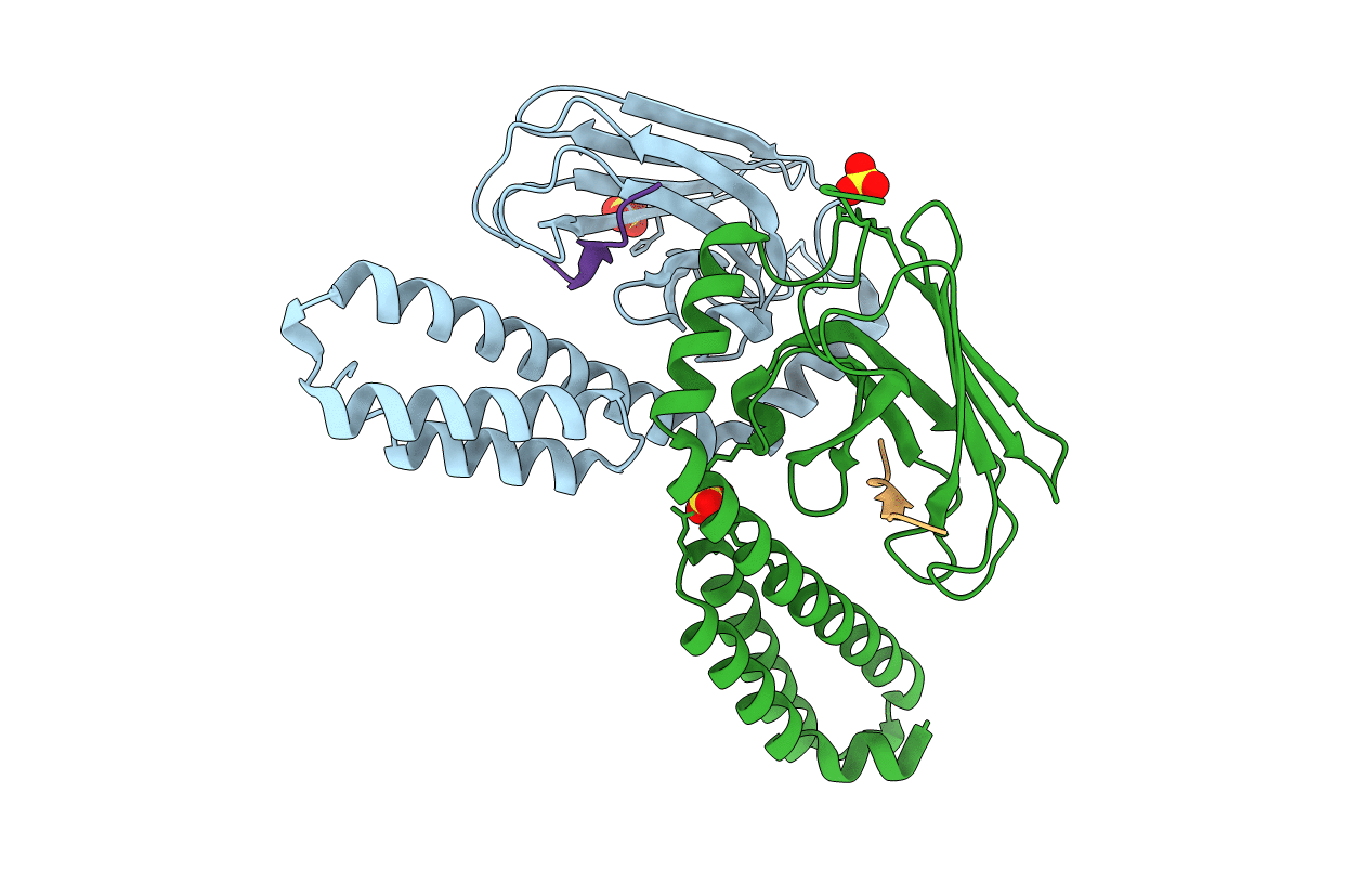
Crystal structure of the substrate binding domain of E.coli DnaK in complex with bovine Bac7(15-28)
Biological Source:
Source Organism:
Escherichia coli (Taxon ID: 83333)
Bos taurus (Taxon ID: 9913)
Host Organism:
Method Details:
Experimental Method:
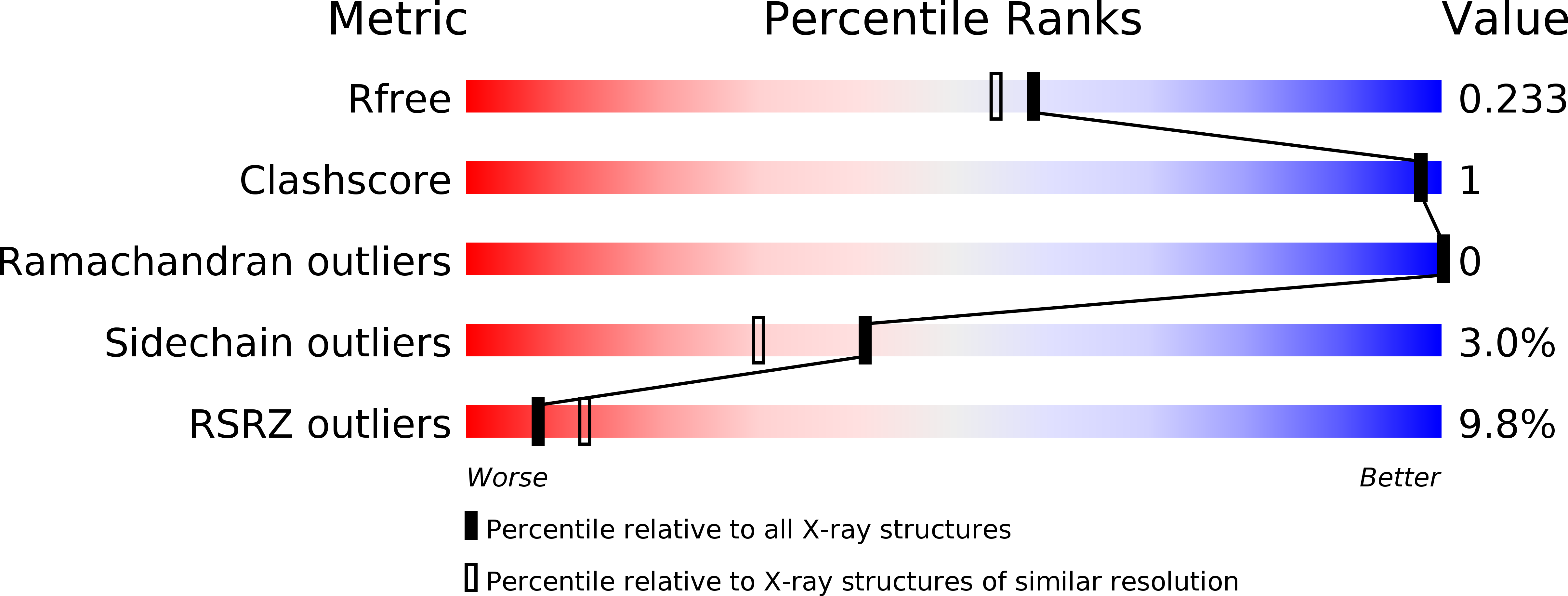
Resolution:
1.95 Å
R-Value Free:
0.21
R-Value Work:
0.18
R-Value Observed:
0.18
Space Group:
P 21 21 2
Macromolecular Entities
Structures with similar UniProt ID
Protein Blast
Polymer Type:polypeptide(L)
Molecule:Chaperone protein DnaK
Gene (Uniprot):dnaK
Chain IDs:A, B
Chain Length:219
Number of Molecules:2
Biological Source:Escherichia coli
Structures with similar UniProt ID
Protein Blast
Polymer Type:polypeptide(L)
Molecule:Cathelicidin-3
Gene (Uniprot):CATHL3
Chain IDs:C, D
Chain Length:14
Number of Molecules:2
Biological Source:Bos taurus
Ligand Molecules
Primary Citation
Structural Identification of DnaK Binding Sites within Bovine and Sheep Bactenecin Bac7.
Protein Pept.Lett. 21 407 412 (2014)
PMID: 24164259

Abstact

Bacterial resistance against common antibiotics is an increasing health problem. New pharmaceuticals for the treatment of infections caused by resistant pathogens are needed. Small proline-rich antimicrobial peptides (PrAMPs) from insects are known to bind intracellularly to the conventional substrate binding cleft of the E. coli Hsp70 chaperone DnaK. Furthermore, bactenecins from mammals, members of the cathelicidin family, also contain potential DnaK binding sites. Crystal structures of bovine and sheep Bac7 in complex with the DnaK substrate binding domain show that the peptides bind in the forward binding mode with a leucine positioned in the central hydrophobic pocket. In most structures, proline and arginine residues preceding leucine occupy the hydrophobic DnaK binding sites -1 and -2. Within bovine Bac7, four potential DnaK binding sites were identified.

Legend

Protein

Chemical

Disease

Primary Citation of related structures