4JSD image
Deposition Date 2013-03-22
Release Date 2013-10-09
Last Version Date 2023-09-20
Entry Detail
PDB ID:
4JSD
Title:
The X-ray crystal structure of a thermophilic cellobiose binding protein bound with laminaribiose
Biological Source:
Source Organism:
Host Organism:
Method Details:
Experimental Method:
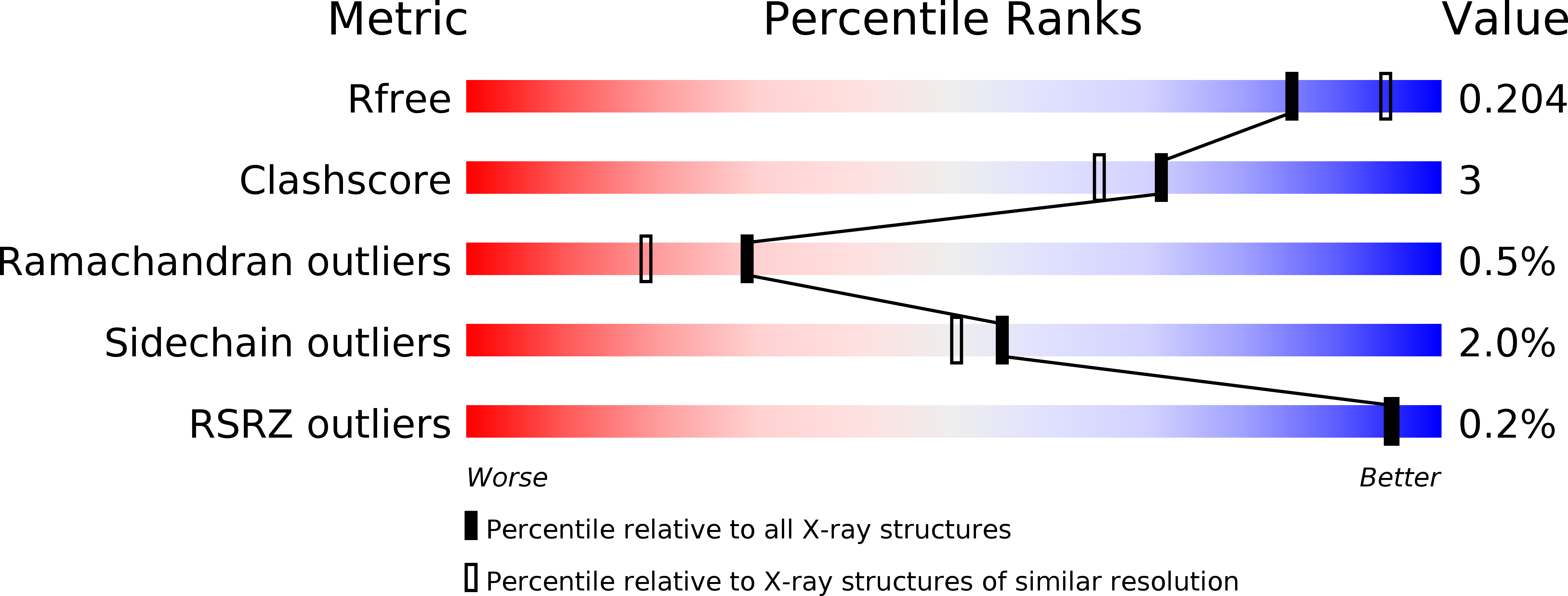
Resolution:
2.05 Å
R-Value Free:
0.20
R-Value Work:
0.18
R-Value Observed:
0.18
Space Group:
P 21 21 21
Macromolecular Entities
Polymer Type:polypeptide(L)
Molecule:Oligopeptide ABC transporter, periplasmic oligopeptide-binding protein
Gene (Uniprot):TM_0031
Chain IDs:A
Chain Length:582
Number of Molecules:1
Biological Source:Thermotoga maritima
Ligand Molecules
Peptide-like Molecules
PRD_900024
Primary Citation
Molecular details of ligand selectivity determinants in a promiscuous beta-glucan periplasmic binding protein.
Bmc Struct.Biol. 13 18 18 (2013)
PMID: 24090243 DOI: 10.1186/1472-6807-13-18

Abstact

BACKGROUND Members of the periplasmic binding protein (PBP) superfamily utilize a highly conserved inter-domain ligand binding site that adapts to specifically bind a chemically diverse range of ligands. This paradigm of PBP ligand binding specificity was recently altered when the structure of the Thermotoga maritima cellobiose-binding protein (tmCBP) was solved. The tmCBP binding site is bipartite, comprising a canonical solvent-excluded region (subsite one), adjacent to a solvent-filled cavity (subsite two) where specific and semi-specific ligand recognition occur, respectively. RESULTS A molecular level understanding of binding pocket adaptation mechanisms that simultaneously allow both ligand specificity at subsite one and promiscuity at subsite two has potentially important implications in ligand binding and drug design studies. We sought to investigate the determinants of ligand binding selectivity in tmCBP through biophysical characterization of tmCBP in the presence of varying β-glucan oligosaccharides. Crystal structures show that whilst the amino acids that comprise both the tmCBP subsite one and subsite two binding sites remain fixed in conformation regardless of which ligands are present, the rich hydrogen bonding potential of water molecules may facilitate the ordering and the plasticity of this unique PBP binding site. CONCLUSIONS The identification of the roles these water molecules play in ligand recognition suggests potential mechanisms that can be utilized to adapt a single ligand binding site to recognize multiple distinct ligands.

Legend

Protein

Chemical

Disease

Primary Citation of related structures