Deposition Date
2013-03-21
Release Date
2013-05-08
Last Version Date
2023-09-20
Entry Detail
PDB ID:
4JRP
Keywords:
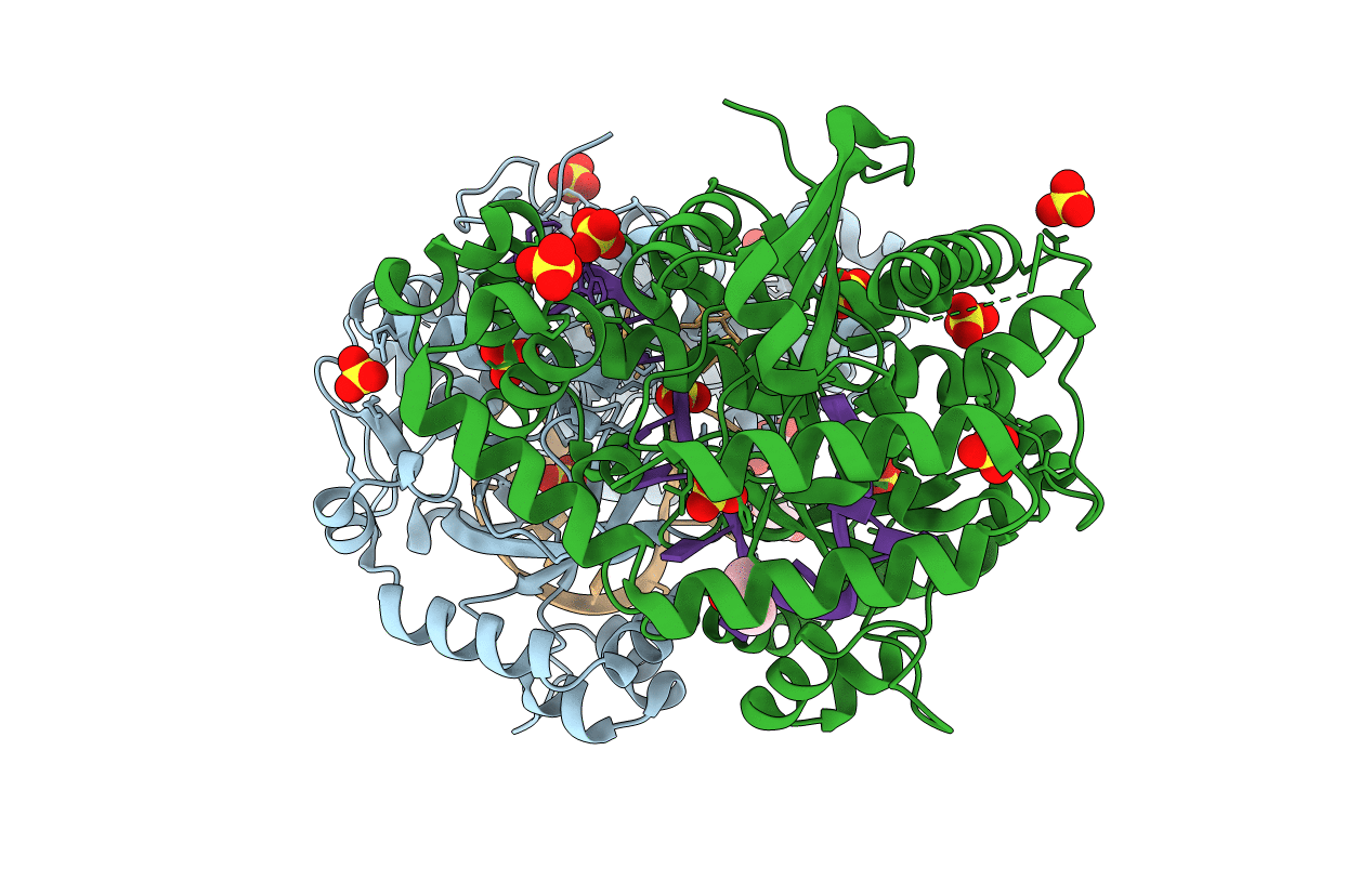
Title:
Structure of E. coli Exonuclease I in complex with a 5cy-dT13 oligonucleotide
Biological Source:
Source Organism(s):
Escherichia coli (Taxon ID: 83333)
Expression System(s):
Method Details:
Experimental Method:
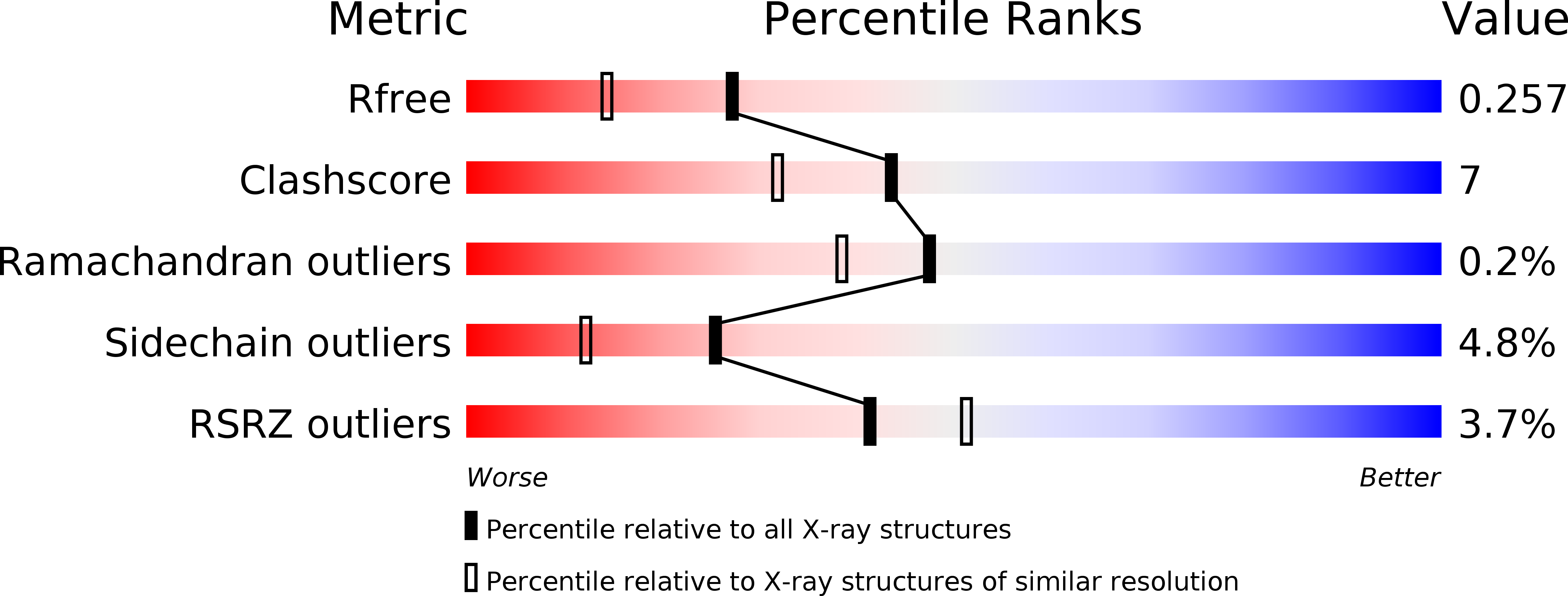
Resolution:
1.95 Å
R-Value Free:
0.25
R-Value Work:
0.21
R-Value Observed:
0.21
Space Group:
P 43