4JRA image
Entry Detail
PDB ID:
4JRA
Keywords:
Title:
CRYSTAL STRUCTURE OF THE BOTULINUM NEUROTOXIN A RECEPTOR-BINDING DOMAIN IN COMPLEX WITH THE LUMINAL DOMAIN Of SV2C
Biological Source:
Host Organism:
PDB Version:
Deposition Date:
2013-03-21
Release Date:
2013-11-20
Method Details:
Experimental Method:
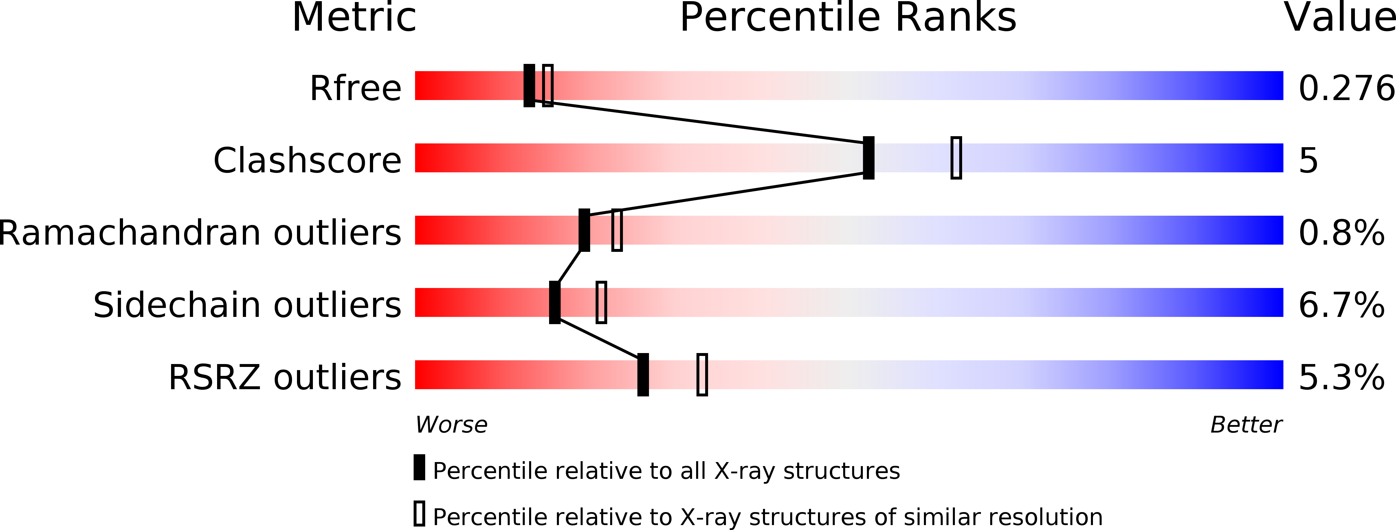
Resolution:
2.30 Å
R-Value Free:
0.26
R-Value Work:
0.23
R-Value Observed:
0.24
Space Group:
C 1 2 1
Macromolecular Entities
Polymer Type:polypeptide(L)
Description:Botulinum neurotoxin type A
Chain IDs:A, B
Chain Length:443
Number of Molecules:2
Biological Source:Clostridium botulinum
Polymer Type:polypeptide(L)
Description:Synaptic vesicle glycoprotein 2C
Chain IDs:C, D
Chain Length:136
Number of Molecules:2
Biological Source:Homo sapiens
Primary Citation
Structural basis for recognition of synaptic vesicle protein 2C by botulinum neurotoxin A.
Nature 505 108 111 (2014)
PMID: 24240280 DOI: 10.1038/nature12732

Abstact

Botulinum neurotoxin A (BoNT/A) belongs to the most dangerous class of bioweapons. Despite this, BoNT/A is used to treat a wide range of common medical conditions such as migraines and a variety of ocular motility and movement disorders. BoNT/A is probably best known for its use as an antiwrinkle agent in cosmetic applications (including Botox and Dysport). BoNT/A application causes long-lasting flaccid paralysis of muscles through inhibiting the release of the neurotransmitter acetylcholine by cleaving synaptosomal-associated protein 25 (SNAP-25) within presynaptic nerve terminals. Two types of BoNT/A receptor have been identified, both of which are required for BoNT/A toxicity and are therefore likely to cooperate with each other: gangliosides and members of the synaptic vesicle glycoprotein 2 (SV2) family, which are putative transporter proteins that are predicted to have 12 transmembrane domains, associate with the receptor-binding domain of the toxin. Recently, fibroblast growth factor receptor 3 (FGFR3) has also been reported to be a potential BoNT/A receptor. In SV2 proteins, the BoNT/A-binding site has been mapped to the luminal domain, but the molecular details of the interaction between BoNT/A and SV2 are unknown. Here we determined the high-resolution crystal structure of the BoNT/A receptor-binding domain (BoNT/A-RBD) in complex with the SV2C luminal domain (SV2C-LD). SV2C-LD consists of a right-handed, quadrilateral β-helix that associates with BoNT/A-RBD mainly through backbone-to-backbone interactions at open β-strand edges, in a manner that resembles the inter-strand interactions in amyloid structures. Competition experiments identified a peptide that inhibits the formation of the complex. Our findings provide a strong platform for the development of novel antitoxin agents and for the rational design of BoNT/A variants with improved therapeutic properties.

Legend

Protein

Chemical

Disease

Primary Citation of related structures