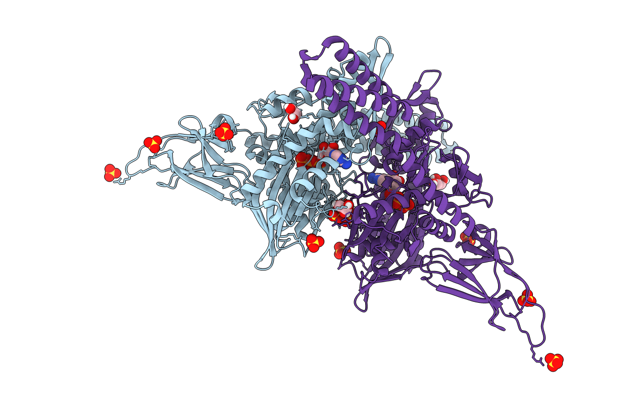
4JNE image
Deposition Date 2013-03-15
Release Date 2013-05-29
Last Version Date 2024-02-28
Entry Detail
PDB ID:
4JNE
Keywords:
Title:
Allosteric opening of the polypeptide-binding site when an Hsp70 binds ATP
Biological Source:
Source Organism:
Host Organism:
Method Details:
Experimental Method:
Resolution:
1.96 Å
R-Value Free:
0.20
R-Value Work:
0.17
R-Value Observed:
0.17
Space Group:
I 4 2 2
Macromolecular Entities
Polymer Type:polypeptide(L)
Molecule:Hsp70 CHAPERONE DnaK
Gene (Uniprot):dnaK
Mutations:N432M,Q433G,S434G
Chain IDs:A, B
Chain Length:608
Number of Molecules:2
Biological Source:Escherichia coli
Primary Citation
Allosteric opening of the polypeptide-binding site when an Hsp70 binds ATP.
Nat.Struct.Mol.Biol. 20 900 907 (2013)
PMID: 23708608 DOI: 10.1038/nsmb.2583

Abstact

The 70-kilodalton (kDa) heat-shock proteins (Hsp70s) are ubiquitous molecular chaperones essential for cellular protein folding and proteostasis. Each Hsp70 has two functional domains: a nucleotide-binding domain (NBD), which binds and hydrolyzes ATP, and a substrate-binding domain (SBD), which binds extended polypeptides. NBD and SBD interact little when in the presence of ADP; however, ATP binding allosterically couples the polypeptide- and ATP-binding sites. ATP binding promotes polypeptide release; polypeptide rebinding stimulates ATP hydrolysis. This allosteric coupling is poorly understood. Here we present the crystal structure of an intact ATP-bound Hsp70 from Escherichia coli at 1.96-Å resolution. The ATP-bound NBD adopts a unique conformation, forming extensive interfaces with an SBD that has changed radically, having its α-helical lid displaced and the polypeptide-binding channel of its β-subdomain restructured. These conformational changes, together with our biochemical assays, provide a structural explanation for allosteric coupling in Hsp70 activity.

Legend

Protein

Chemical

Disease

Primary Citation of related structures