4JLC image
Deposition Date 2013-03-12
Release Date 2013-06-19
Last Version Date 2023-09-20
Entry Detail
PDB ID:
4JLC
Title:
Crystal structure of mouse TBK1 bound to SU6668
Biological Source:
Source Organism:
Mus musculus (Taxon ID: 10090)
Host Organism:
Method Details:
Experimental Method:
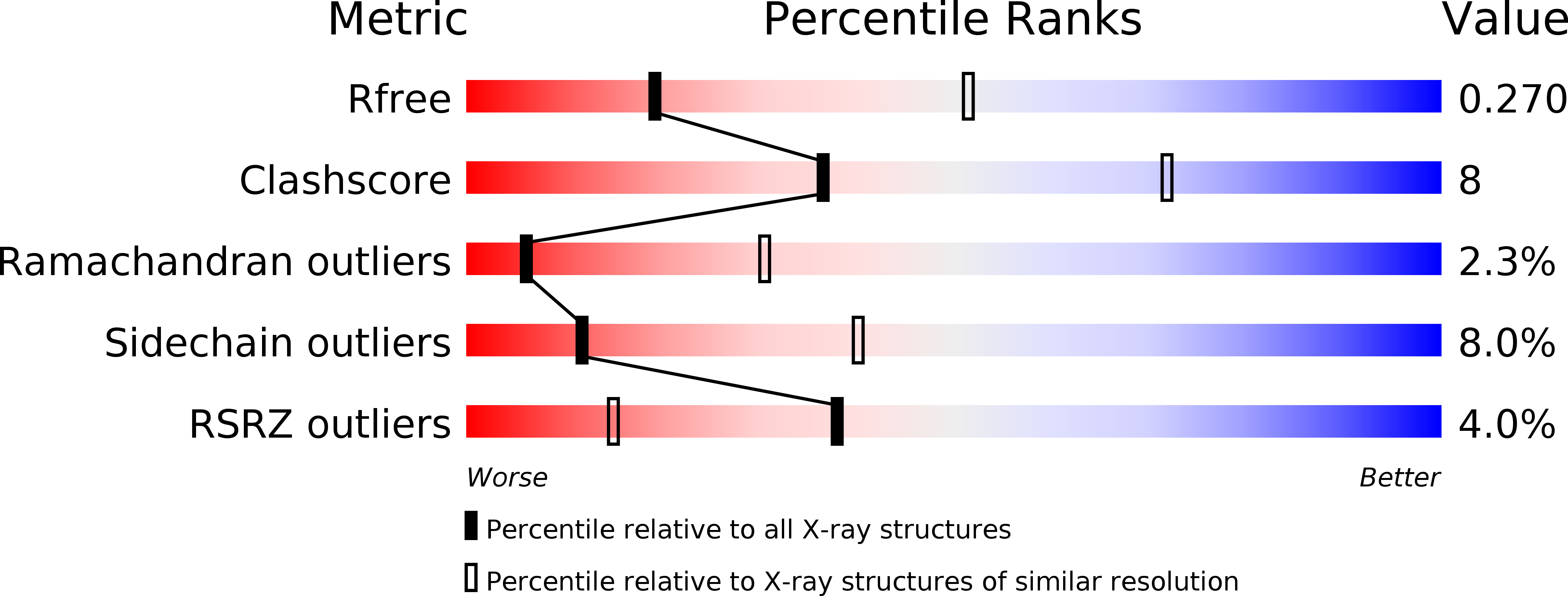
Resolution:
3.00 Å
R-Value Free:
0.26
R-Value Work:
0.23
R-Value Observed:
0.23
Space Group:
P 32 2 1
Macromolecular Entities
Polymer Type:polypeptide(L)
Molecule:Serine/threonine-protein kinase TBK1
Gene (Uniprot):Tbk1
Chain IDs:A
Chain Length:657
Number of Molecules:1
Biological Source:Mus musculus
Ligand Molecules
Primary Citation
Structural Insights into the Functions of TBK1 in Innate Antimicrobial Immunity.
Structure 21 1137 1148 (2013)
PMID: 23746807 DOI: 10.1016/j.str.2013.04.025

Abstact

Tank-binding kinase 1 (TBK1) is a serine/threonine protein-kinase mediating innate antimicrobial immunity. TBK1 is involved in the signaling of TLRs, RLRs, and STING-mediated sensing of cytosolic DNA. Stimulation of these receptors results in the activation of TBK1, which phosphorylates interferon regulatory factor (IRF)-3. Phosphorylated IRF-3 translocates into the nucleus to initiate the transcription of the interferon (IFN)-β gene. Here, we show that TBK1 is activated by autophosphorylation at residue Ser172. Structures of TBK1 bound to two inhibitors showed that TBK1 has the IκB kinase fold with three distinct domains: the kinase domain, the ubiquitin-like domain, and the scaffold and dimerization domain. However, the overall structures of the TBK1 monomer and its dimer are different from IKKβ in the arrangements of the three domains and in dimer formation. Phosphorylation of IRF-3 by TBK1 in vitro results in its oligomerization, and phosphorylation of residue Ser386 plays a key role in IRF-3 activation.

Legend

Protein

Chemical

Disease

Primary Citation of related structures