4JKQ image
Deposition Date 2013-03-11
Release Date 2014-04-02
Last Version Date 2023-09-20
Entry Detail
PDB ID:
4JKQ
Title:
Crystal structure of the N-terminal region of the human ryanodine receptor 2
Biological Source:
Source Organism:
Homo sapiens (Taxon ID: 9606)
Host Organism:
Method Details:
Experimental Method:
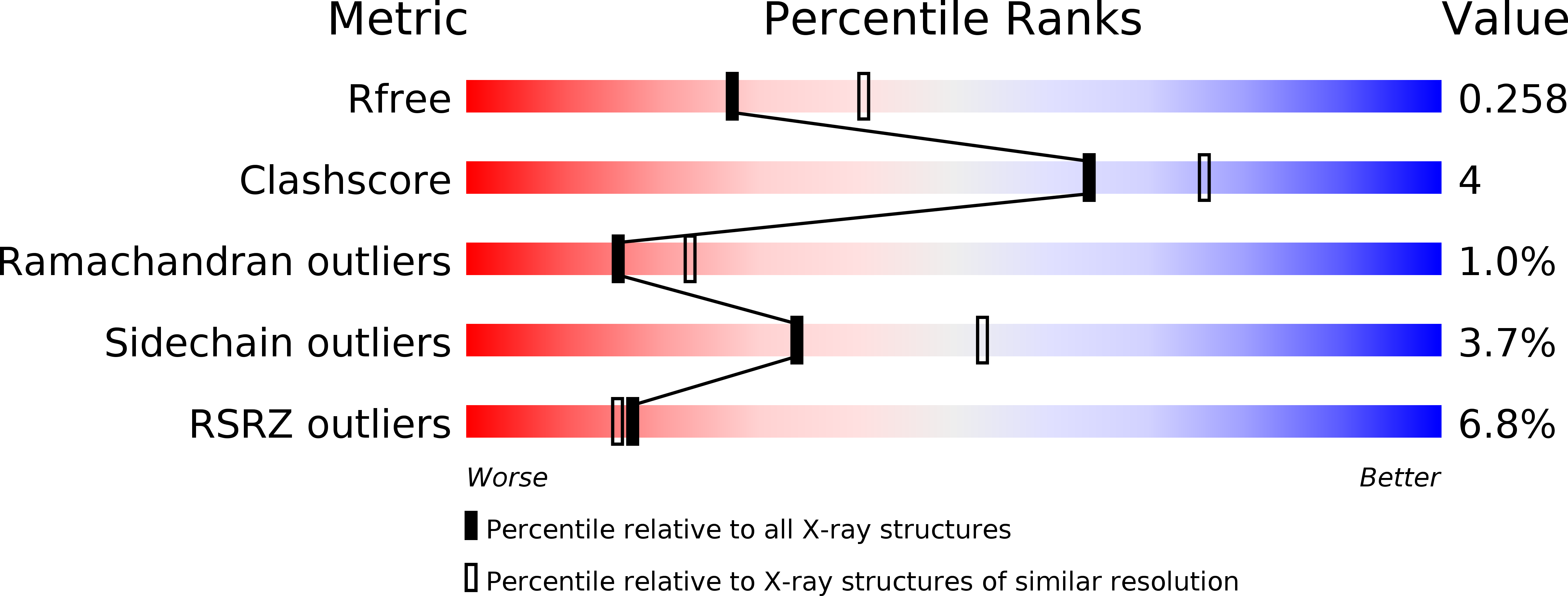
Resolution:
2.39 Å
R-Value Free:
0.26
R-Value Work:
0.22
R-Value Observed:
0.22
Space Group:
P 42 21 2
Macromolecular Entities
Polymer Type:polypeptide(L)
Molecule:Ryanodine receptor 2
Gene (Uniprot):RYR2
Chain IDs:A
Chain Length:606
Number of Molecules:1
Biological Source:Homo sapiens
Primary Citation
Structural insights into the human RyR2 N-terminal region involved in cardiac arrhythmias.
Acta Crystallogr.,Sect.D 70 2897 2912 (2014)
PMID: 25372681 DOI: 10.1107/S1399004714020343

Abstact

Human ryanodine receptor 2 (hRyR2) mediates calcium release from the sarcoplasmic reticulum, enabling cardiomyocyte contraction. The N-terminal region of hRyR2 (amino acids 1-606) is the target of >30 arrhythmogenic mutations and contains a binding site for phosphoprotein phosphatase 1. Here, the solution and crystal structures determined under near-physiological conditions, as well as a homology model of the hRyR2 N-terminal region, are presented. The N-terminus is held together by a unique network of interactions among its three domains, A, B and C, in which the central helix (amino acids 410-437) plays a prominent stabilizing role. Importantly, the anion-binding site reported for the mouse RyR2 N-terminal region is notably absent from the human RyR2. The structure concurs with the differential stability of arrhythmogenic mutations in the central helix (R420W, I419F and I419F/R420W) which are owing to disparities in the propensity of mutated residues to form energetically favourable or unfavourable contacts. In solution, the N-terminus adopts a globular shape with a prominent tail that is likely to involve residues 545-606, which are unresolved in the crystal structure. Docking the N-terminal domains into cryo-electron microscopy maps of the closed and open RyR1 conformations reveals C(α) atom movements of up to 8 Å upon channel gating, and predicts the location of the leucine-isoleucine zipper segment and the interaction site for spinophilin and phosphoprotein phosphatase 1 on the RyR surface.

Legend

Protein

Chemical

Disease

Primary Citation of related structures