
Deposition Date
2013-03-07
Release Date
2014-03-12
Last Version Date
2025-07-23
Entry Detail
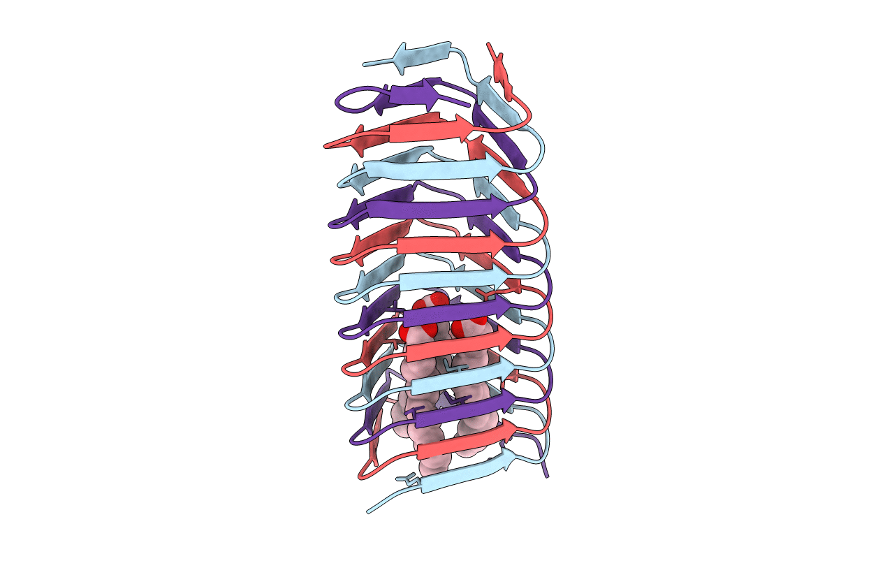
PDB ID:
4JJ2
Keywords:
Title:
High resolution structure of a C-terminal fragment of the T4 phage gp5 beta-helix
Biological Source:
Source Organism(s):
Enterobacteria phage T4 (Taxon ID: 10665)
Expression System(s):
Method Details:
Experimental Method:
Resolution:
1.28 Å
R-Value Free:
0.17
R-Value Work:
0.15
R-Value Observed:
0.15
Space Group:
C 2 2 21
