4JH1 image
Entry Detail
PDB ID:
4JH1
Keywords:
Title:
Crystal Structure of FosB from Bacillus cereus with Zinc and Sulfate at 1.55 A Resolution -SAD Phasing
Biological Source:
Source Organism:
Host Organism:
PDB Version:
Deposition Date:
2013-03-04
Release Date:
2013-10-02
Method Details:
Experimental Method:
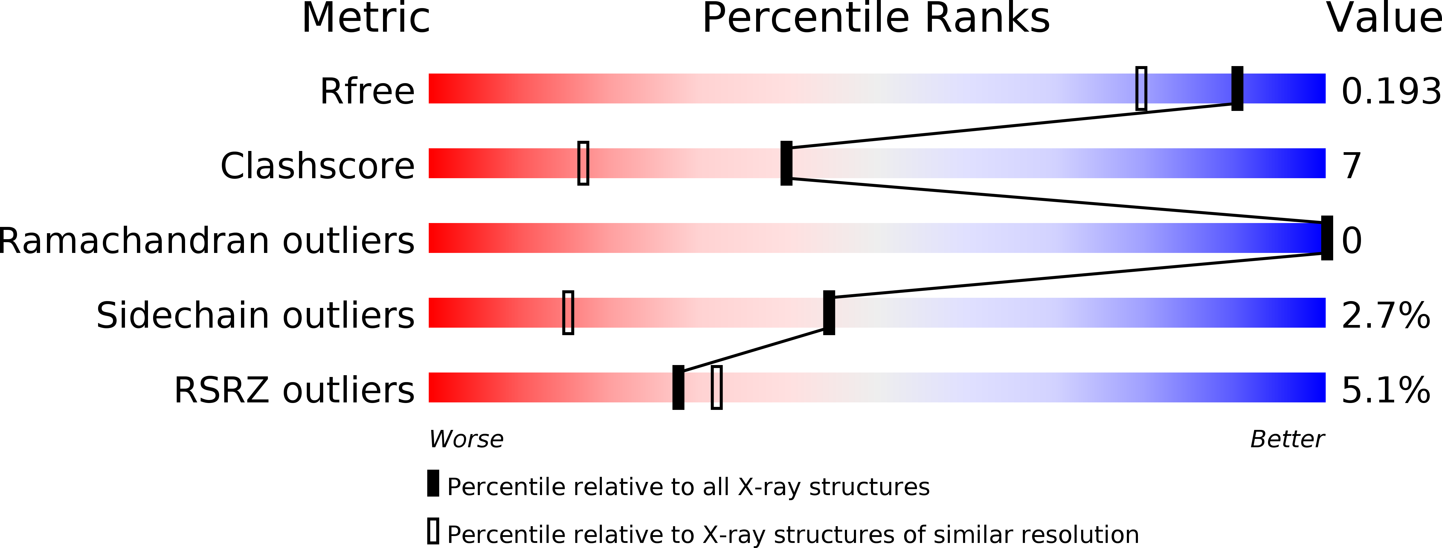
Resolution:
1.55 Å
R-Value Free:
0.18
R-Value Work:
0.13
R-Value Observed:
0.13
Space Group:
P 21 21 21
Macromolecular Entities
Polymer Type:polypeptide(L)
Description:Metallothiol transferase FosB
Chain IDs:A, B
Chain Length:138
Number of Molecules:2
Biological Source:Bacillus cereus
Primary Citation
Structural and Chemical Aspects of Resistance to the Antibiotic Fosfomycin Conferred by FosB from Bacillus cereus.
Biochemistry 52 7350 7362 (2013)
PMID: 24004181 DOI: 10.1021/bi4009648

Abstact

The fosfomycin resistance enzymes, FosB, from Gram-positive organisms, are M(2+)-dependent thiol tranferases that catalyze nucleophilic addition of either L-cysteine (L-Cys) or bacillithiol (BSH) to the antibiotic, resulting in a modified compound with no bacteriacidal properties. Here we report the structural and functional characterization of FosB from Bacillus cereus (FosB(Bc)). The overall structure of FosB(Bc), at 1.27 Å resolution, reveals that the enzyme belongs to the vicinal oxygen chelate (VOC) superfamily. Crystal structures of FosB(Bc) cocrystallized with fosfomycin and a variety of divalent metals, including Ni(2+), Mn(2+), Co(2+), and Zn(2+), indicate that the antibiotic coordinates to the active site metal center in an orientation similar to that found in the structurally homologous manganese-dependent fosfomycin resistance enzyme, FosA. Surface analysis of the FosB(Bc) structures show a well-defined binding pocket and an access channel to C1 of fosfomycin, the carbon to which nucleophilic addition of the thiol occurs. The pocket and access channel are appropriate in size and shape to accommodate L-Cys or BSH. Further investigation of the structures revealed that the fosfomycin molecule, anchored by the metal, is surrounded by a cage of amino acids that hold the antibiotic in an orientation such that C1 is centered at the end of the solvent channel, positioning the compound for direct nucleophilic attack by the thiol substrate. In addition, the structures of FosB(Bc) in complex with the L-Cys-fosfomycin product (1.55 Å resolution) and in complex with the bacillithiol-fosfomycin product (1.77 Å resolution) coordinated to a Mn(2+) metal in the active site have been determined. The L-Cys moiety of either product is located in the solvent channel, where the thiol has added to the backside of fosfomycin C1 located at the end of the channel. Concomitant kinetic analyses of FosB(Bc) indicated that the enzyme has a preference for BSH over L-Cys when activated by Mn(2+) and is inhibited by Zn(2+). The fact that Zn(2+) is an inhibitor of FosB(Bc) was used to obtain a ternary complex structure of the enzyme with both fosfomycin and L-Cys bound.

Legend

Protein

Chemical

Disease

Primary Citation of related structures