4JGF image
Deposition Date 2013-03-01
Release Date 2013-05-22
Last Version Date 2024-02-28
Entry Detail
PDB ID:
4JGF
Title:
Crystal Structure of the Cataract-Causing P23T gamma D-Crystallin Mutant
Biological Source:
Source Organism:
Homo sapiens (Taxon ID: 9606)
Host Organism:
Method Details:
Experimental Method:
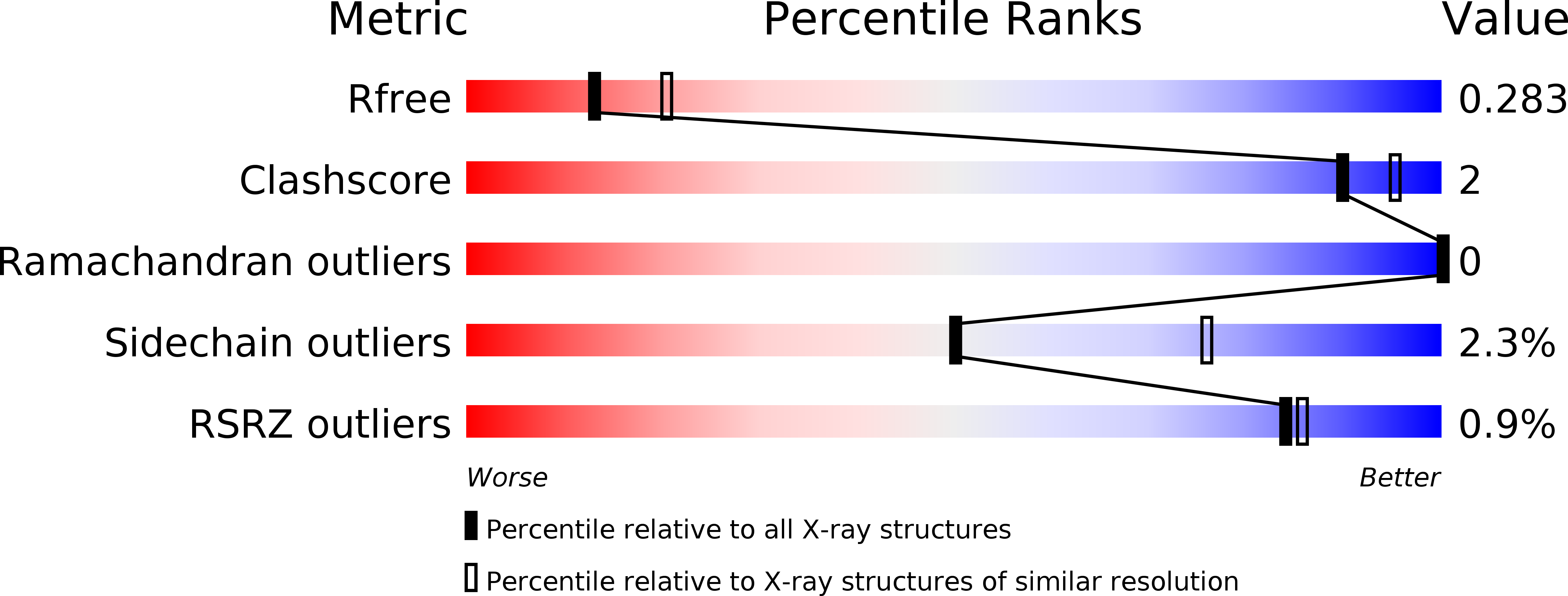
Resolution:
2.50 Å
R-Value Free:
0.28
R-Value Work:
0.22
R-Value Observed:
0.23
Space Group:
P 1 21 1
Macromolecular Entities
Polymer Type:polypeptide(L)
Molecule:Gamma-crystallin D
Gene (Uniprot):CRYGD
Mutations:P23T
Chain IDs:A, B
Chain Length:171
Number of Molecules:2
Biological Source:Homo sapiens
Primary Citation
Crystal structure of the cataract-causing P23T gamma D-crystallin mutant.
Proteins 81 1493 1498 (2013)
PMID: 23670788 DOI: 10.1002/prot.24321

Abstact

Up to now, efforts to crystallize the cataract-associated P23T mutant of human γD-crystallin have not been successful. Therefore, insights into the light scattering mechanism of this mutant have been exclusively obtained from solution work. Here we present the first crystal structure of the P23T mutant at 2.5 Å resolution. The protein exhibits essentially the same overall structure as seen for the wild-type protein. Based on our structural data, we confirm that no major conformational changes are caused by the mutation, and that solution phase properties of the mutant appear exclusively associated with cataract formation.

Legend

Protein

Chemical

Disease

Primary Citation of related structures