4JDR image
Deposition Date 2013-02-25
Release Date 2013-04-24
Last Version Date 2024-11-06
Entry Detail
PDB ID:
4JDR
Keywords:
Title:
Dihydrolipoamide dehydrogenase of pyruvate dehydrogenase from escherichia coli
Biological Source:
Source Organism:
Escherichia coli (Taxon ID: 562)
Host Organism:
Method Details:
Experimental Method:
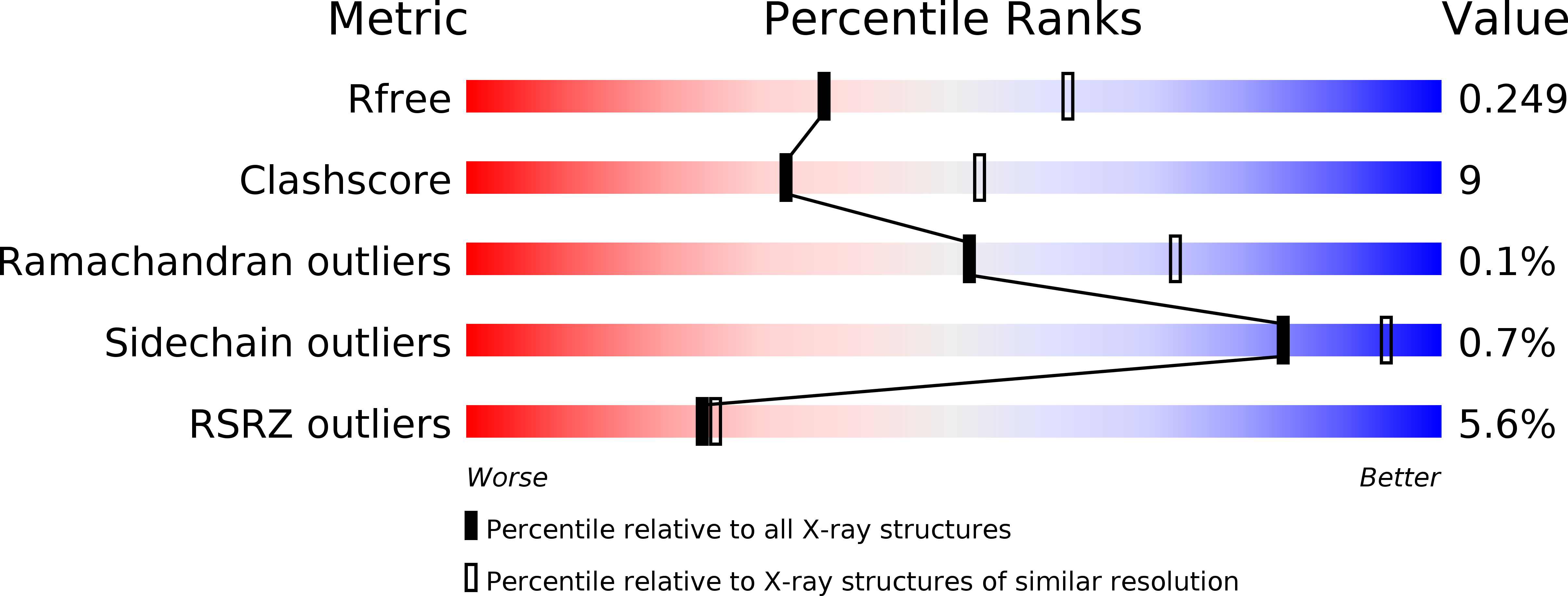
Resolution:
2.50 Å
R-Value Free:
0.25
R-Value Work:
0.21
R-Value Observed:
0.21
Space Group:
I 4 2 2
Macromolecular Entities
Structures with similar UniProt ID
Protein Blast
Polymer Type:polypeptide(L)
Molecule:Dihydrolipoyl dehydrogenase
Gene (Uniprot):lpdA
Chain IDs:A, B
Chain Length:474
Number of Molecules:2
Biological Source:Escherichia coli
Primary Citation
Insight to the Interaction of the Dihydrolipoamide Acetyltransferase (E2) Core with the Peripheral Components in the Escherichia coli Pyruvate Dehydrogenase Complex via Multifaceted Structural Approaches.
J.Biol.Chem. 288 15402 15417 (2013)
PMID: 23580650 DOI: 10.1074/jbc.M113.466789

Abstact

Multifaceted structural approaches were undertaken to investigate interaction of the E2 component with E3 and E1 components from the Escherichia coli pyruvate dehydrogenase multienzyme complex (PDHc), as a representative of the PDHc from Gram-negative bacteria. The crystal structure of E3 at 2.5 Å resolution reveals similarity to other E3 structures and was an important starting point for understanding interaction surfaces between E3 and E2. Biochemical studies revealed that R129E-E2 and R150E-E2 substitutions in the peripheral subunit-binding domain (PSBD) of E2 greatly diminished PDHc activity, affected interactions with E3 and E1 components, and affected reductive acetylation of E2. Because crystal structures are unavailable for any complete E2-containing complexes, peptide-specific hydrogen/deuterium exchange mass spectrometry was used to identify loci of interactions between 3-lipoyl E2 and E3. Two peptides from the PSBD, including Arg-129, and three peptides from E3 displayed statistically significant reductions in deuterium uptake resulting from interaction between E3 and E2. Of the peptides identified on E3, two were from the catalytic site, and the third was from the interface domain, which for all known E3 structures is believed to interact with the PSBD. NMR clearly demonstrates that there is no change in the lipoyl domain structure on complexation with E3. This is the first instance where the entire wild-type E2 component was employed to understand interactions with E3. A model for PSBD-E3 binding was independently constructed and found to be consistent with the importance of Arg-129, as well as revealing other electrostatic interactions likely stabilizing this complex.

Legend

Protein

Chemical

Disease

Primary Citation of related structures