
Deposition Date
2013-02-22
Release Date
2013-08-07
Last Version Date
2023-11-08
Entry Detail
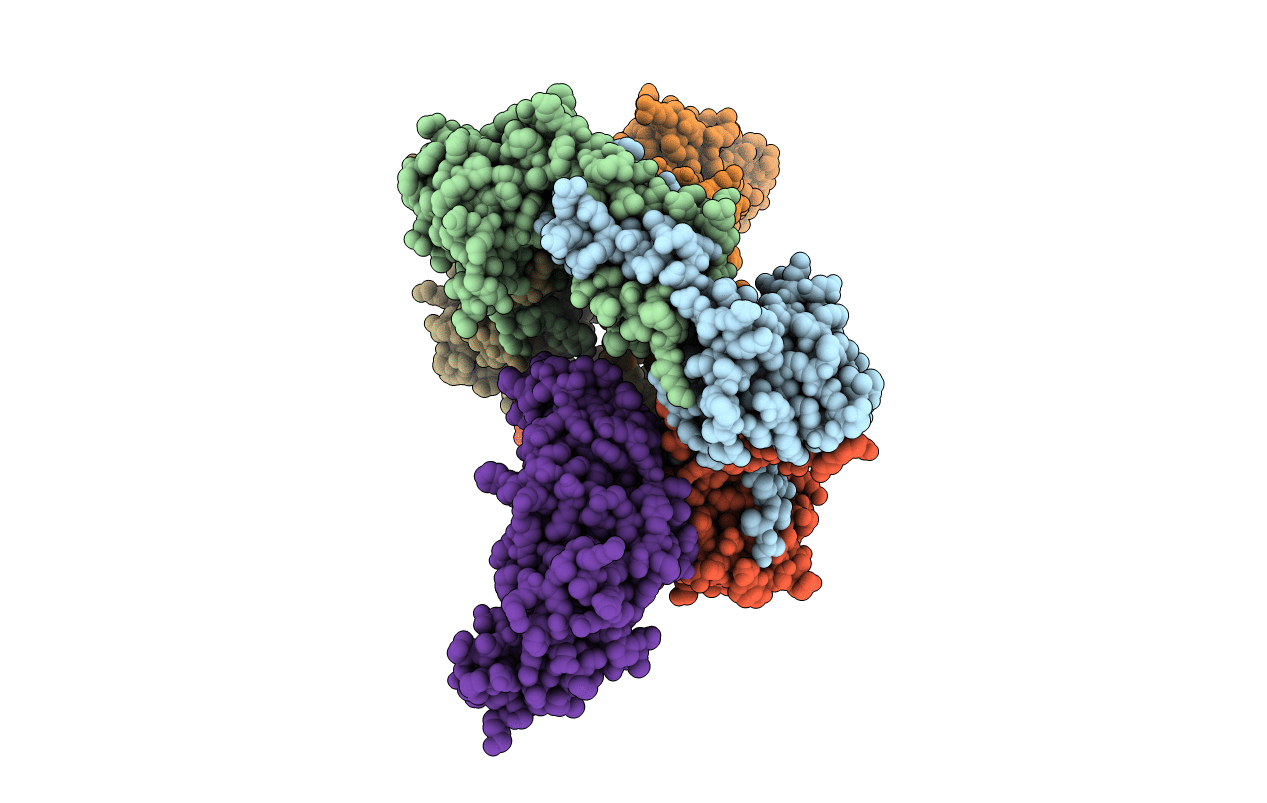
PDB ID:
4JCV
Keywords:
Title:
Crystal structure of the RecOR complex in an open conformation
Biological Source:
Source Organism(s):
Deinococcus radiodurans (Taxon ID: 243230)
Expression System(s):
Method Details:
Experimental Method:
Resolution:
3.34 Å
R-Value Free:
0.27
R-Value Work:
0.24
R-Value Observed:
0.24
Space Group:
P 1
