Deposition Date
2013-02-20
Release Date
2013-05-08
Last Version Date
2023-09-20
Entry Detail
PDB ID:
4JBU
Keywords:
Title:
1.65A structure of the T3SS tip protein LcrV (G28-D322, C273S) from Yersinia pestis
Biological Source:
Source Organism(s):
Yersinia pestis (Taxon ID: 632)
Expression System(s):
Method Details:
Experimental Method:
Resolution:
1.65 Å
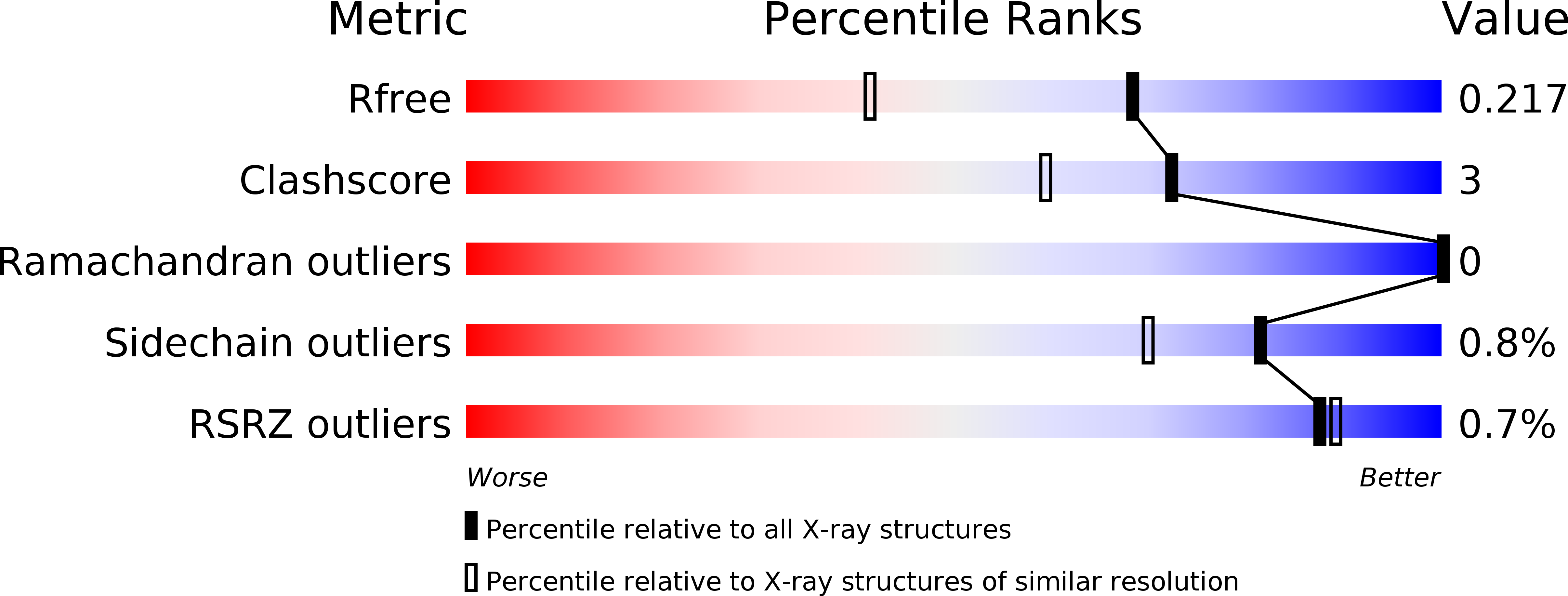
R-Value Free:
0.22
R-Value Work:
0.17
R-Value Observed:
0.17
Space Group:
P 1 21 1