
Deposition Date
2013-02-20
Release Date
2013-12-11
Last Version Date
2024-10-30
Entry Detail
PDB ID:
4JBS
Keywords:
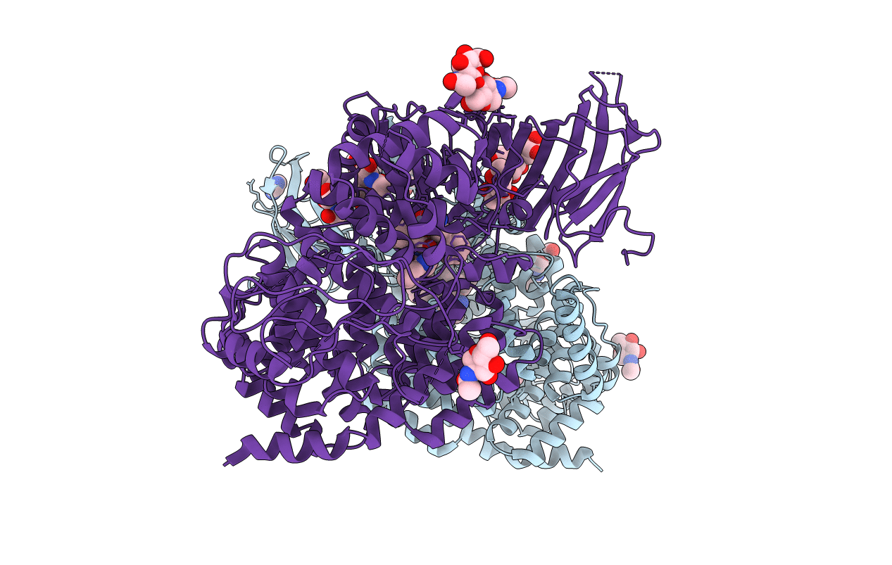
Title:
Crystal structure of the human Endoplasmic Reticulum Aminopeptidase 2 in complex with PHOSPHINIC PSEUDOTRIPEPTIDE inhibitor.
Biological Source:
Source Organism(s):
Homo sapiens (Taxon ID: 9606)
Expression System(s):
Method Details:
Experimental Method:
Resolution:
2.79 Å
R-Value Free:
0.27
R-Value Work:
0.20
R-Value Observed:
0.21
Space Group:
P 1 21 1
