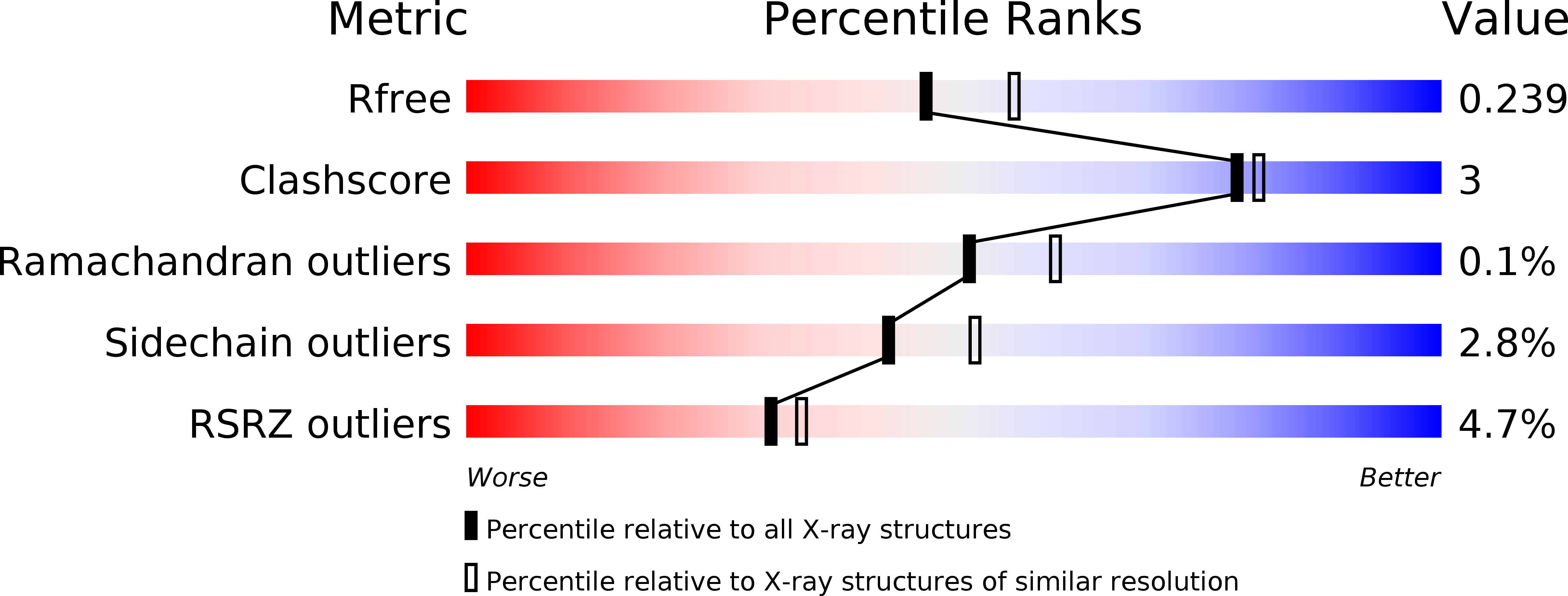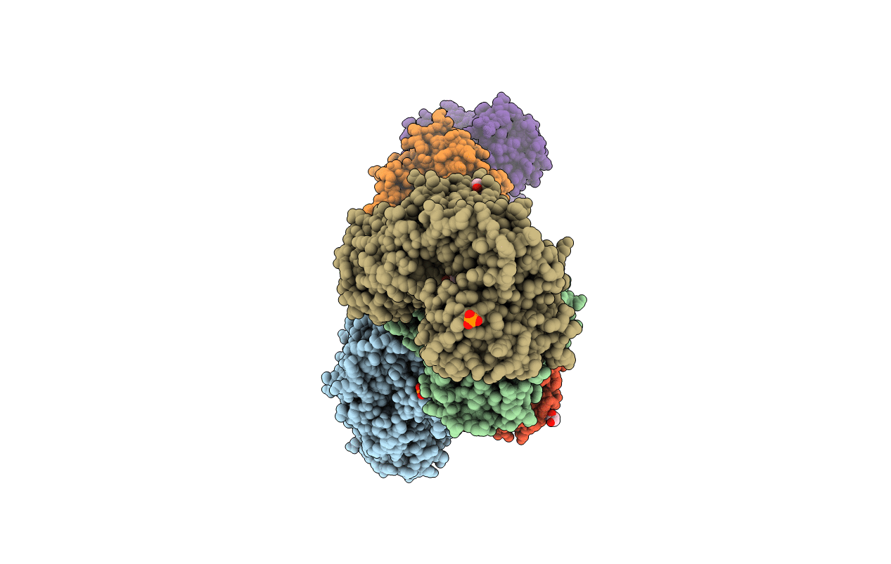
4JAX image
Deposition Date 2013-02-19
Release Date 2013-05-01
Last Version Date 2024-10-09
Entry Detail
PDB ID:
4JAX
Keywords:
Title:
Crystal structure of dimeric KlHxk1 in crystal form X
Biological Source:
Source Organism:
Host Organism:
Method Details:
Experimental Method:
Resolution:
2.26 Å
R-Value Free:
0.23
R-Value Work:
0.20
R-Value Observed:
0.20
Space Group:
P 21 21 21
Macromolecular Entities
Polymer Type:polypeptide(L)
Molecule:Hexokinase
Gene (Uniprot):RAG5
Chain IDs:A, B, C, D, E, F
Chain Length:485
Number of Molecules:6
Biological Source:Kluyveromyces lactis
Modified Residue
Compound ID Chain ID Parent Comp ID Details 2D Image
SEP A SER PHOSPHOSERINE
Primary Citation
In vivo phosphorylation and in vitro autophosphorylation-inactivation of Kluyveromyces lactis hexokinase KlHxk1.
Biochem.Biophys.Res.Commun. 435 313 318 (2013)
PMID: 23583397 DOI: 10.1016/j.bbrc.2013.03.121

Abstact

The bifunctional hexokinase KlHxk1 is a key component of glucose-dependent signal transduction in Kluyveromyces lactis. KlHxk1 is phosphorylated in vivo and undergoes ATP-dependent autophosphorylation-inactivation in vitro. This study identifies serine-15 as the site of in vivo phosphorylation and serine-157 as the autophosphorylation-inactivation site. X-ray crystallography of the in vivo phosphorylated enzyme indicates the existence of a ring-shaped symmetrical homodimer carrying two phosphoserine-15 residues. In contrast, small-angle X-ray scattering and equilibrium sedimentation analyses reveal the existence of monomeric phosphoserine-15 KlHxk1 in solution. While phosphorylation at serine-15 and concomitant homodimer dissociation are likely to be involved in glucose signalling, mechanism and putative physiological significance of KlHxk1 inactivation by autophosphorylation at serine-157 remain to be established.

Legend

Protein

Chemical

Disease

Primary Citation of related structures