4J7H image
Deposition Date 2013-02-13
Release Date 2013-05-22
Last Version Date 2024-11-27
Entry Detail
PDB ID:
4J7H
Title:
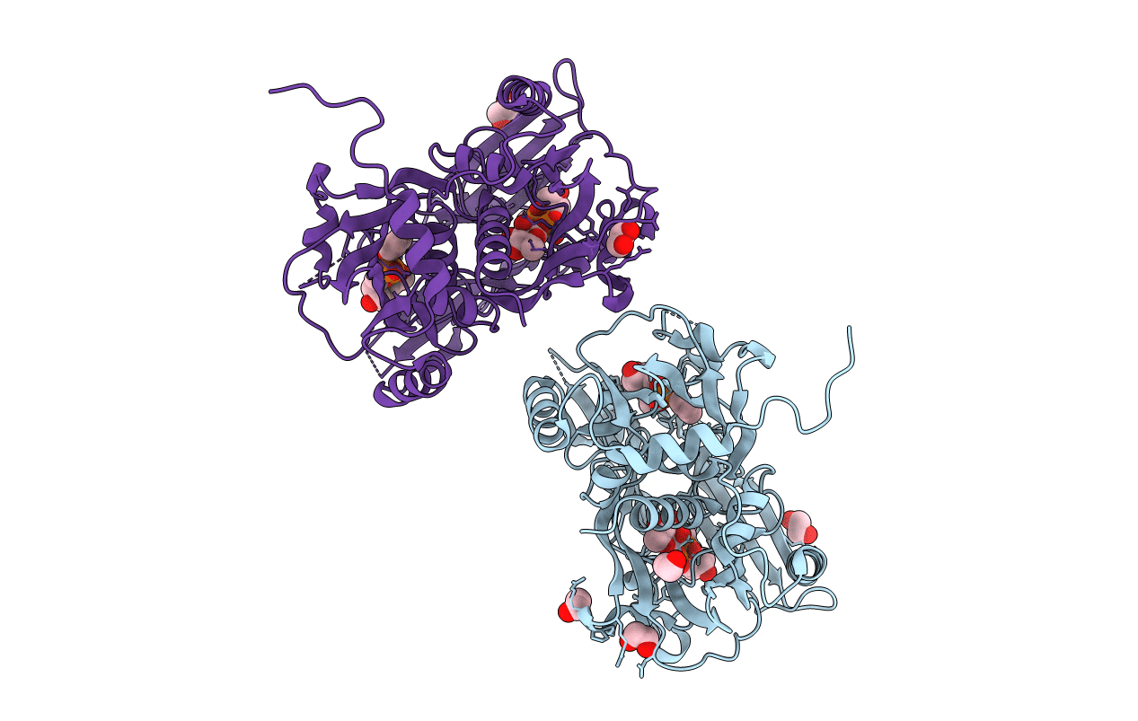
Crystal structure of EvaA, a 2,3-dehydratase in complex with dTDP-benzene and dTDP-rhamnose
Biological Source:
Source Organism:
Host Organism:
Method Details:
Experimental Method:
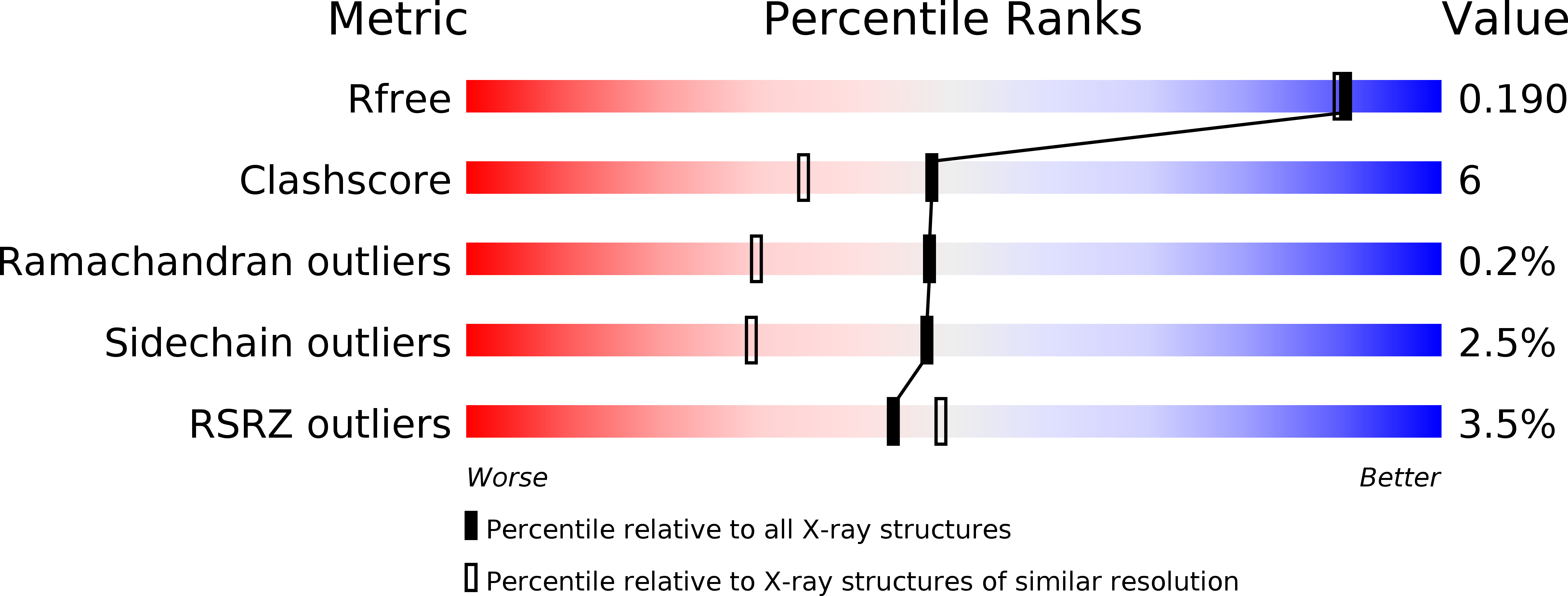
Resolution:
1.69 Å
R-Value Free:
0.19
R-Value Work:
0.16
R-Value Observed:
0.16
Space Group:
P 21 21 2
Macromolecular Entities
Structures with similar UniProt ID
Protein Blast
Polymer Type:polypeptide(L)
Molecule:EvaA 2,3-dehydratase
Gene (Uniprot):evaA
Mutagens:R381A
Chain IDs:A, B
Chain Length:471
Number of Molecules:2
Biological Source:Amycolatopsis orientalis
Primary Citation
Structure of EvaA: A Paradigm for Sugar 2,3-Dehydratases.
Biochemistry 52 2078 2088 (2013)
PMID: 23473392 DOI: 10.1021/bi400176n

Abstact

Unusual deoxysugars found appended to natural products often provide or enhance the pharmacokinetic activities of the parent compound. The preferred carbohydrate donors for the biosynthesis of such glycosylated natural products are the dTDP-linked sugars. Many of the biologically relevant dTDP-deoxysugars are constructed around the 2,6-dideoxyhexoses or the 2,3(4),6-trideoxyhexoses. A key step in the biosynthesis of these sugars is the removal of the hexose C-2' hydroxyl group and the oxidation of the C-3' hydroxyl group to a carbonyl moiety. Enzymes that catalyze these reactions are referred to as 2,3-dehydratases and have been, for the most part, largely uncharacterized. Here we report the first structural analysis of a sugar 2,3-dehydratase. For this investigation, the enzyme, EvaA, was cloned from Amycolatopsis orientalis, and the structure was solved and refined to a nominal resolution of 1.7 Å. On the basis of the resulting model, it is clear that EvaA belongs to the large Nudix hydrolase superfamily and is most similar to GDP-mannose hydrolase. Each subunit of the EvaA dimer folds into two domains that clearly arose via gene duplication. Two dTDP-sugar binding pockets, A and B, are present in each EvaA subunit. On the basis of site-directed mutagenesis experiments and activity assays, it appears that pocket A functions as the active site and pocket B is simply a remnant left behind from the gene duplication event. As 2,3-dehydration is crucial for the biosynthesis of many unusual deoxysugars, this investigation provides key structural insight into this widely conserved reaction.

Legend

Protein

Chemical

Disease

Primary Citation of related structures