4J6S image
Deposition Date 2013-02-11
Release Date 2013-10-02
Last Version Date 2024-11-20
Entry Detail
PDB ID:
4J6S
Keywords:
Title:
14-3-3gamma complexed with the N-terminal sequence of tyrosine hydroxylase (residues 1-43)
Biological Source:
Source Organism:
Homo sapiens (Taxon ID: 9606)
Host Organism:
Method Details:
Experimental Method:
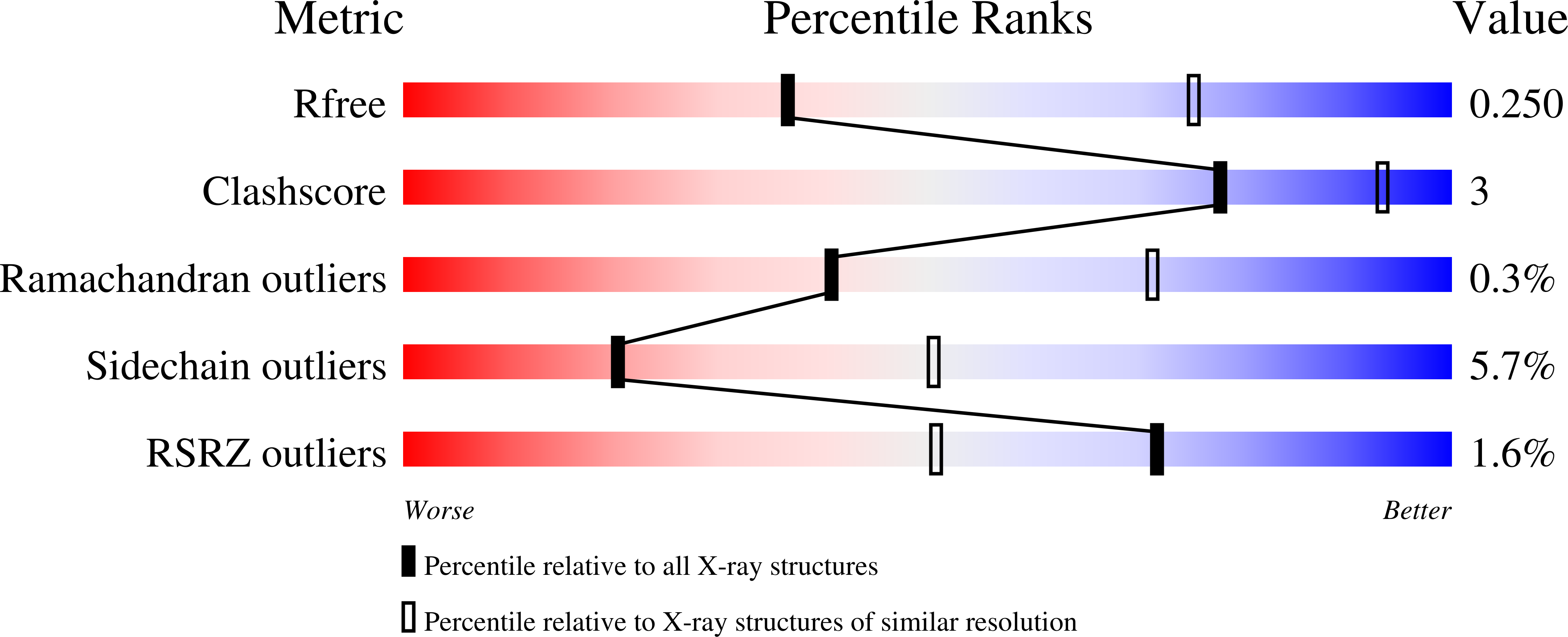
Resolution:
3.08 Å
R-Value Free:
0.25
R-Value Work:
0.21
R-Value Observed:
0.21
Space Group:
P 21 21 21
Macromolecular Entities
Polymer Type:polypeptide(L)
Molecule:14-3-3 protein gamma
Gene (Uniprot):YWHAG
Chain IDs:A, B, C, D
Chain Length:255
Number of Molecules:4
Biological Source:Homo sapiens
Polymer Type:polypeptide(L)
Molecule:N-terminal motif of tyrosine hydroxylase
Chain IDs:E, F, G, H
Chain Length:43
Number of Molecules:4
Biological Source:Homo sapiens
Modified Residue
Compound ID Chain ID Parent Comp ID Details 2D Image
SEP E SER PHOSPHOSERINE
Ligand Molecules
Primary Citation
The N-terminal sequence of tyrosine hydroxylase is a conformationally versatile motif that binds 14-3-3 proteins and membranes.
J.Mol.Biol. 426 150 168 (2014)
PMID: 24055376 DOI: 10.1016/j.jmb.2013.09.012

Abstact

Tyrosine hydroxylase (TH) catalyzes the rate-limiting step in the synthesis of catecholamine neurotransmitters, and a reduction in TH activity is associated with several neurological diseases. Human TH is regulated, among other mechanisms, by Ser19-phosphorylation-dependent interaction with 14-3-3 proteins. The N-terminal sequence (residues 1-43), which corresponds to an extension to the TH regulatory domain, also interacts with negatively charged membranes. By using X-ray crystallography together with molecular dynamics simulations and structural bioinformatics analysis, we have probed the conformations of the Ser19-phosphorylated N-terminal peptide [THp-(1-43)] bound to 14-3-3γ, free in solution and bound to a phospholipid bilayer, and of the unphosphorylated peptide TH-(1-43) both free and bilayer bound. As seen in the crystal structure of THp-(1-43) complexed with 14-3-3γ, the region surrounding pSer19 adopts an extended conformation in the bound state, whereas THp-(1-43) adopts a bent conformation when free in solution, with higher content of secondary structure and higher number of internal hydrogen bonds. TH-(1-43) in solution presents the highest mobility and least defined structure of all forms studied, and it shows an energetically more favorable interaction with membranes relative to THp-(1-43). Cationic residues, notably Arg15 and Arg16, which are the recognition sites of the kinases phosphorylating at Ser19, are also contributing to the interaction with the membrane. Our results reveal the structural flexibility of this region of TH, in accordance with the functional versatility and conformational adaptation to different partners. Furthermore, this structural information has potential relevance for the development of therapeutics for neurodegenerative disorders, through modulation of TH-partner interactions.

Legend

Protein

Chemical

Disease

Primary Citation of related structures