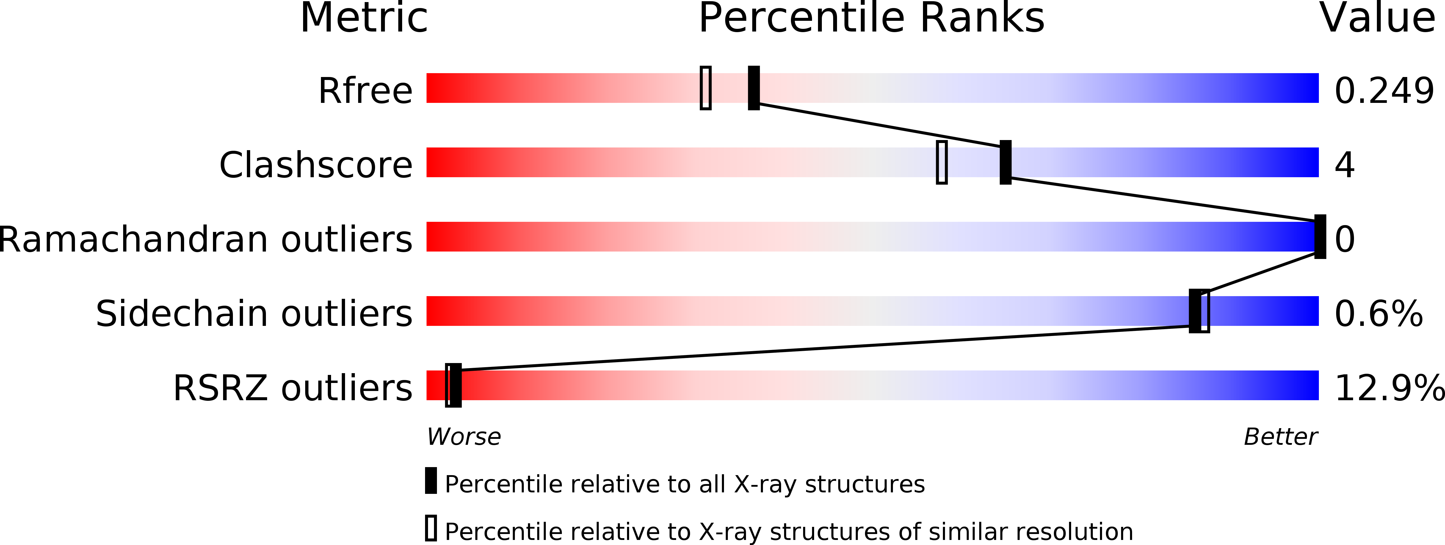
4J5T image
Deposition Date 2013-02-09
Release Date 2013-04-03
Last Version Date 2024-11-20
Entry Detail
PDB ID:
4J5T
Keywords:
Title:
Crystal structure of Processing alpha-Glucosidase I
Biological Source:
Source Organism:
Host Organism:
Method Details:
Experimental Method:
Resolution:
2.04 Å
R-Value Free:
0.23
R-Value Work:
0.22
R-Value Observed:
0.22
Space Group:
P 21 21 21
Macromolecular Entities
Polymer Type:polypeptide(L)
Molecule:Mannosyl-oligosaccharide glucosidase
Gene (Uniprot):CWH41
Chain IDs:A
Chain Length:811
Number of Molecules:1
Biological Source:Saccharomyces cerevisiae
Modified Residue
Compound ID Chain ID Parent Comp ID Details 2D Image
ASN A ASN GLYCOSYLATION SITE
Primary Citation
Specificity of processing alpha-glucosidase I is guided by the substrate conformation: crystallographic and in silico studies.
J.Biol.Chem. 288 13563 13574 (2013)
PMID: 23536181 DOI: 10.1074/jbc.M113.460436

Abstact

BACKGROUND The enzyme “GluI” is key to the synthesis of critical glycoproteins in the cell. RESULTS We have determined the structure of GluI, and modeled binding with its unique sugar substrate. CONCLUSION The specificity of this interaction derives from a unique conformation of the substrate. SIGNIFICANCE Understanding the mechanism of the enzyme is of basic importance and relevant to potential development of antiviral inhibitors. Processing α-glucosidase I (GluI) is a key member of the eukaryotic N-glycosylation processing pathway, selectively catalyzing the first glycoprotein trimming step in the endoplasmic reticulum. Inhibition of GluI activity impacts the infectivity of enveloped viruses; however, despite interest in this protein from a structural, enzymatic, and therapeutic standpoint, little is known about its structure and enzymatic mechanism in catalysis of the unique glycan substrate Glc3Man9GlcNAc2. The first structural model of eukaryotic GluI is here presented at 2-Å resolution. Two catalytic residues are proposed, mutations of which result in catalytically inactive, properly folded protein. Using Autodocking methods with the known substrate and inhibitors as ligands, including a novel inhibitor characterized in this work, the active site of GluI was mapped. From these results, a model of substrate binding has been formulated, which is most likely conserved in mammalian GluI.

Legend

Protein

Chemical

Disease

Primary Citation of related structures