
Deposition Date
2013-02-06
Release Date
2013-11-27
Last Version Date
2024-10-09
Entry Detail
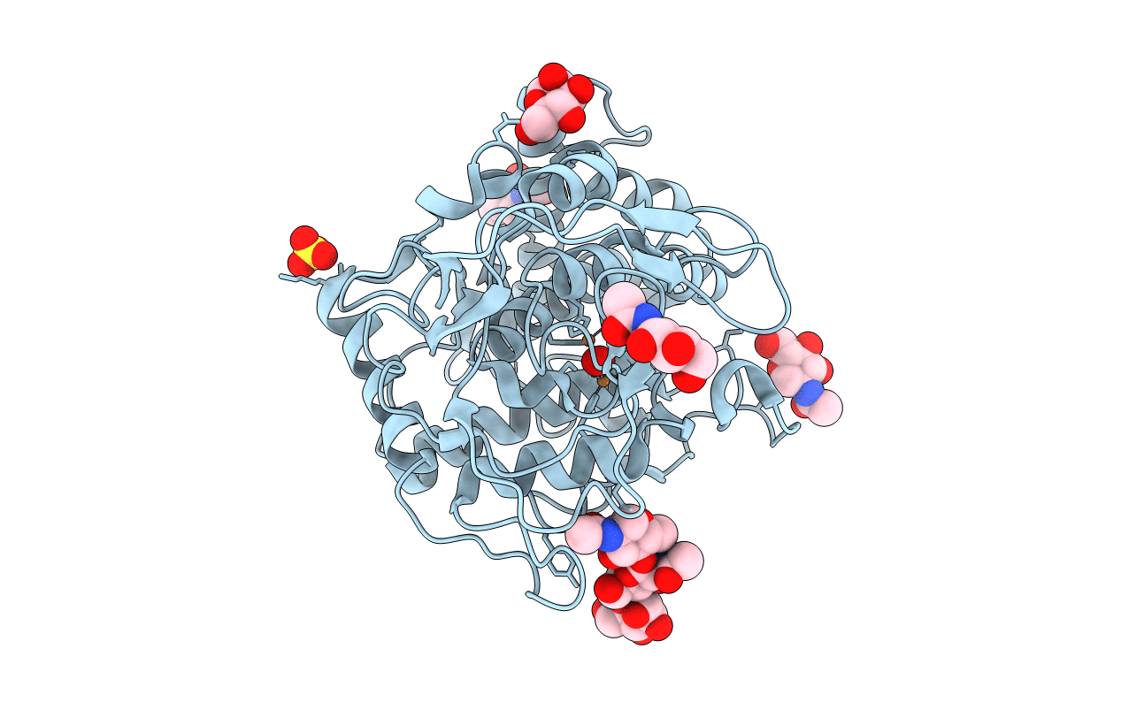
PDB ID:
4J3R
Keywords:
Title:
Crystal structure of catechol oxidase from Aspergillus oryzae, soaked in 4-tert-butylcatechol
Biological Source:
Source Organism(s):
Aspergillus oryzae (Taxon ID: 5062)
Expression System(s):
Method Details:
Experimental Method:
Resolution:
2.20 Å
R-Value Free:
0.22
R-Value Work:
0.18
R-Value Observed:
0.19
Space Group:
P 32 2 1
