Deposition Date
2013-02-05
Release Date
2013-07-24
Last Version Date
2024-11-06
Entry Detail
PDB ID:
4J2V
Keywords:
Title:
Crystal Structure of Equine Serum Albumin in complex with 3,5-diiodosalicylic acid
Biological Source:
Source Organism(s):
Equus caballus (Taxon ID: 9796)
Method Details:
Experimental Method:
Resolution:
2.12 Å
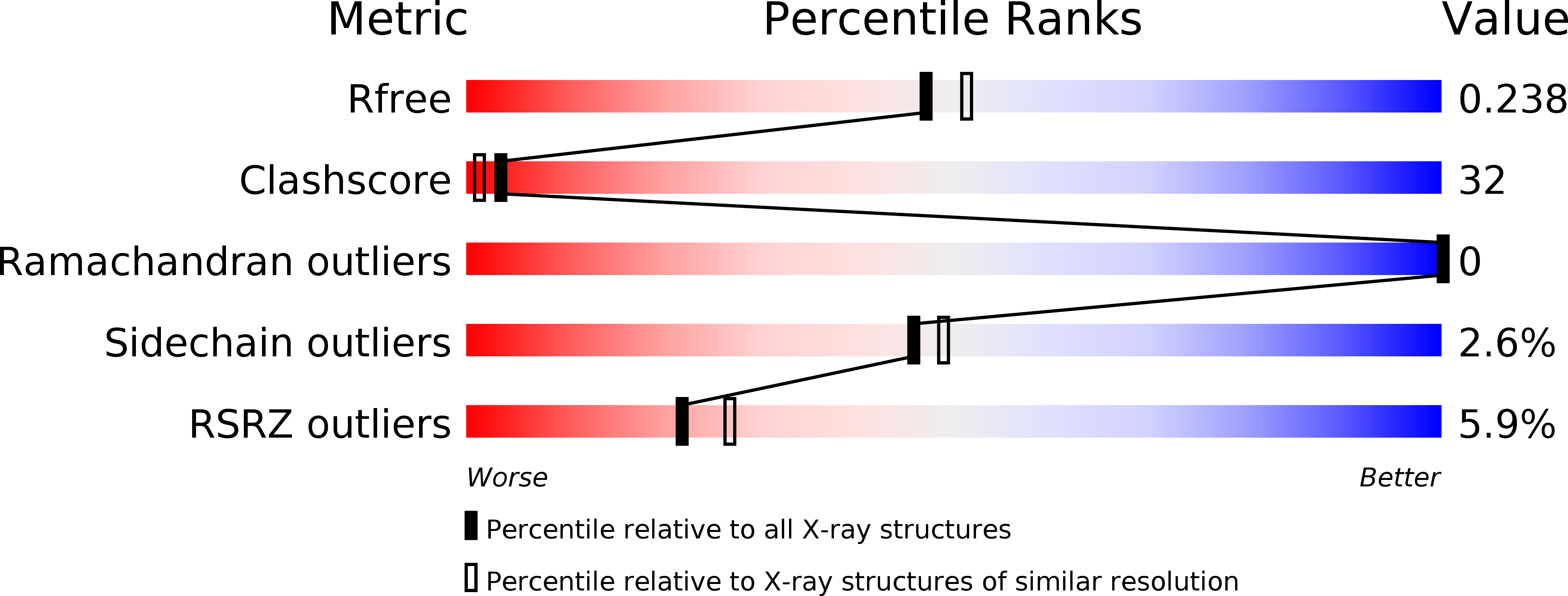
R-Value Free:
0.23
R-Value Work:
0.18
R-Value Observed:
0.18
Space Group:
P 61