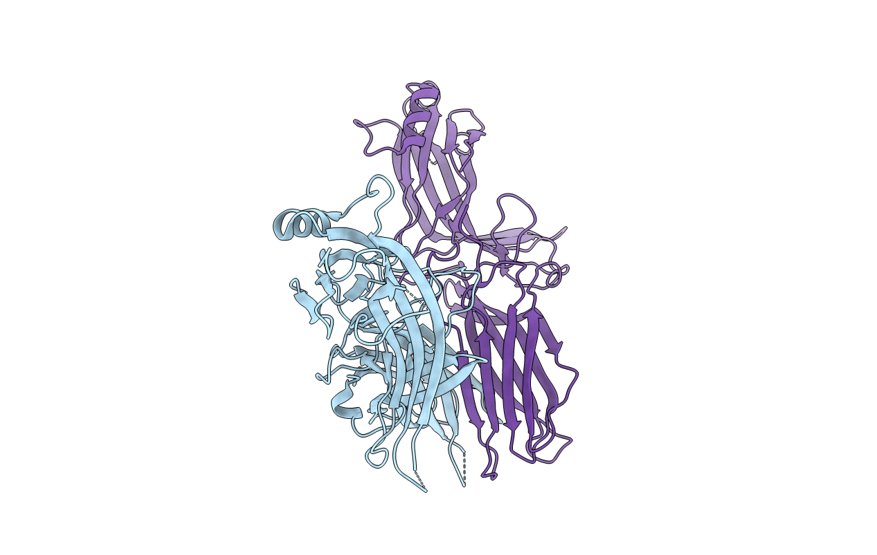
4J2Q image
Entry Detail
PDB ID:
4J2Q
Title:
Crystal structure of C-terminally truncated arrestin reveals mechanism of arrestin activation
Biological Source:
Source Organism:
Host Organism:
PDB Version:
Deposition Date:
2013-02-05
Release Date:
2013-05-01
Method Details:
Experimental Method:
Resolution:
3.00 Å
R-Value Free:
0.27
R-Value Work:
0.24
R-Value Observed:
0.24
Space Group:
P 61 2 2
Macromolecular Entities
Polymer Type:polypeptide(L)
Description:S-arrestin
Chain IDs:A, B
Chain Length:380
Number of Molecules:2
Biological Source:Bos taurus
Primary Citation
Crystal structure of pre-activated arrestin p44.
Nature 497 142 146 (2013)
PMID: 23604253 DOI: 10.1038/nature12133

Abstact

Arrestins interact with G-protein-coupled receptors (GPCRs) to block interaction with G proteins and initiate G-protein-independent signalling. Arrestins have a bi-lobed structure that is stabilized by a long carboxy-terminal tail (C-tail), and displacement of the C-tail by receptor-attached phosphates activates arrestins for binding active GPCRs. Structures of the inactive state of arrestin are available, but it is not known how C-tail displacement activates arrestin for receptor coupling. Here we present a 3.0 Å crystal structure of the bovine arrestin-1 splice variant p44, in which the activation step is mimicked by C-tail truncation. The structure of this pre-activated arrestin is profoundly different from the basal state and gives insight into the activation mechanism. p44 displays breakage of the central polar core and other interlobe hydrogen-bond networks, leading to a ∼21° rotation of the two lobes as compared to basal arrestin-1. Rearrangements in key receptor-binding loops in the central crest region include the finger loop, loop 139 (refs 8, 10, 11) and the sequence Asp 296-Asn 305 (or gate loop), here identified as controlling the polar core. We verified the role of these conformational alterations in arrestin activation and receptor binding by site-directed fluorescence spectroscopy. The data indicate a mechanism for arrestin activation in which C-tail displacement releases critical central-crest loops from restricted to extended receptor-interacting conformations. In parallel, increased flexibility between the two lobes facilitates a proper fitting of arrestin to the active receptor surface. Our results provide a snapshot of an arrestin ready to bind the active receptor, and give an insight into the role of naturally occurring truncated arrestins in the visual system.

Legend

Protein

Chemical

Disease

Primary Citation of related structures