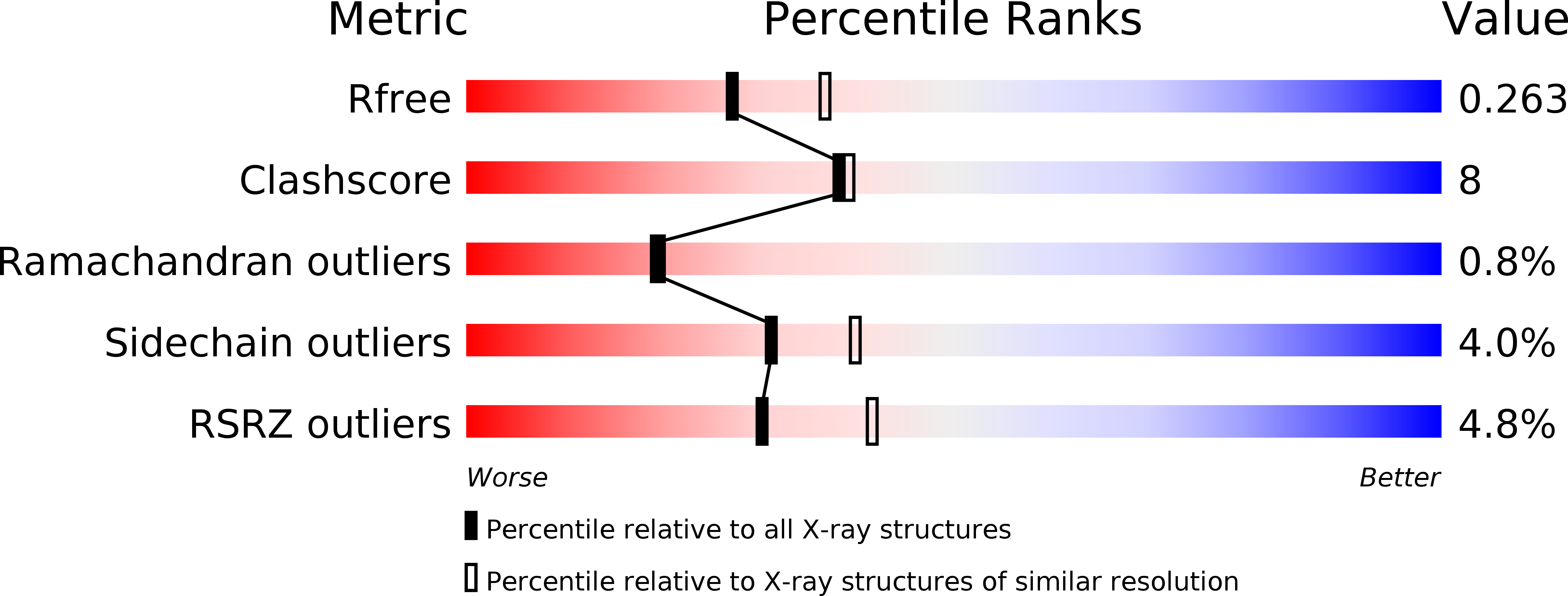
4J2N image
Deposition Date 2013-02-04
Release Date 2013-10-23
Last Version Date 2024-02-28
Entry Detail
PDB ID:
4J2N
Keywords:
Title:
Crystal Structure of mycobacteriophage Pukovnik Xis
Biological Source:
Source Organism:
Host Organism:
Method Details:
Experimental Method:
Resolution:
2.35 Å
R-Value Free:
0.26
R-Value Work:
0.22
R-Value Observed:
0.22
Space Group:
C 2 2 21
Macromolecular Entities
Structures with similar UniProt ID
Protein Blast
Polymer Type:polypeptide(L)
Molecule:Gp37
Gene (Uniprot):37
Chain IDs:A, B, C (auth: D), D (auth: C), E
Chain Length:58
Number of Molecules:5
Biological Source:Mycobacterium phage Pukovnik
Ligand Molecules
Primary Citation
The Structure of Xis Reveals the Basis for Filament Formation and Insight into DNA Bending within a Mycobacteriophage Intasome.
J.Mol.Biol. 426 412 422 (2014)
PMID: 24112940 DOI: 10.1016/j.jmb.2013.10.002

Abstact

The recombination directionality factor, Xis, is a DNA bending protein that determines the outcome of integrase-mediated site-specific recombination by redesign of higher-order protein-DNA architectures. Although the attachment site DNA of mycobacteriophage Pukovnik is likely to contain four sites for Xis binding, Xis crystals contain five subunits in the asymmetric unit, four of which align into a Xis filament and a fifth that is generated by an unusual domain swap. Extensive intersubunit contacts stabilize a bent filament-like arrangement with Xis monomers aligned head to tail. The structure implies a DNA bend of ~120°, which is in agreement with DNA bending measured in vitro. Formation of attR-containing intasomes requires only Int and Xis, distinguishing Pukovnik from lambda. Therefore, we conclude that, in Pukovnik, Xis-induced DNA bending is sufficient to promote intramolecular Int-mediated bridges during intasome formation.

Legend

Protein

Chemical

Disease

Primary Citation of related structures