4J20 image
Deposition Date 2013-02-04
Release Date 2013-10-02
Last Version Date 2023-11-08
Entry Detail
PDB ID:
4J20
Keywords:
Title:
X-ray structure of the cytochrome c-554 from chlorobaculum tepidum
Biological Source:
Source Organism:
Host Organism:
Method Details:
Experimental Method:
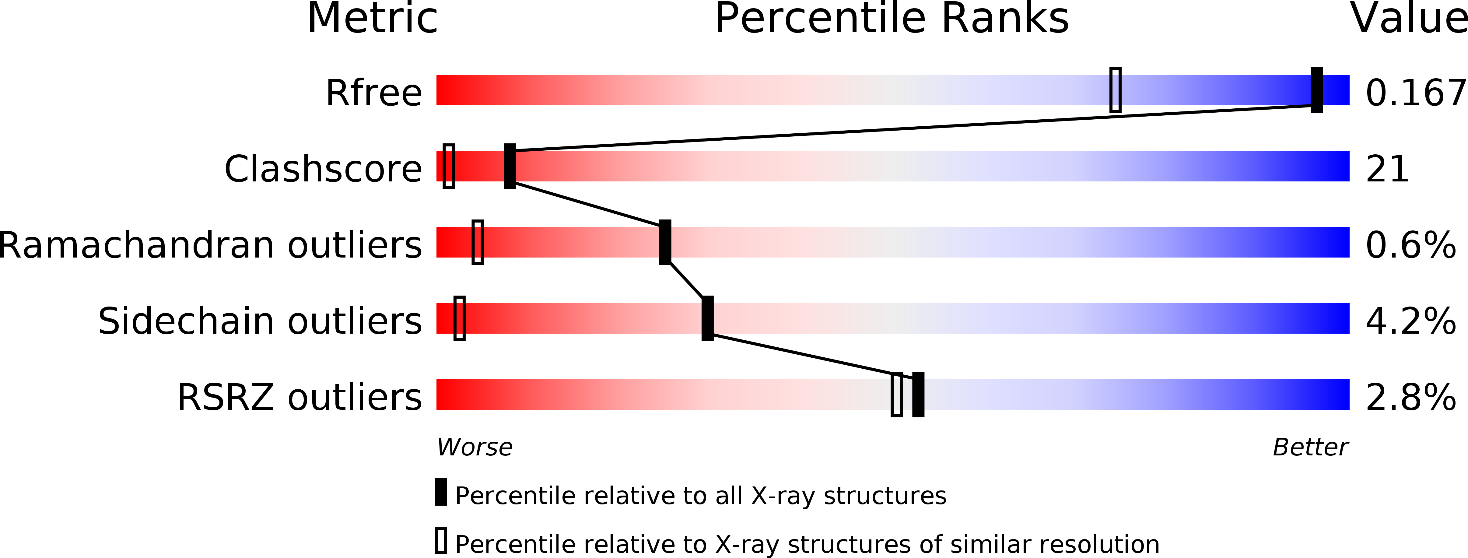
Resolution:
1.30 Å
R-Value Free:
0.16
R-Value Work:
0.15
R-Value Observed:
0.15
Space Group:
C 1 2 1
Macromolecular Entities
Structures with similar UniProt ID
Protein Blast
Polymer Type:polypeptide(L)
Molecule:Cytochrome c-555
Gene (Uniprot):CT0075
Mutagens:P1S, A2T
Chain IDs:A, B
Chain Length:88
Number of Molecules:2
Biological Source:Chlorobium tepidum
Primary Citation
Structure analysis and characterization of the cytochrome c-554 from thermophilic green sulfur photosynthetic bacterium Chlorobaculum tepidum
Photosynth.Res. 118 249 258 (2013)
PMID: 24052268 DOI: 10.1007/s11120-013-9922-2

Abstact

The cytochrome (Cyt) c-554 in thermophilic green photosynthetic bacterium Chlorobaculum tepidum serves as an intermediate electron carrier, transferring electrons to the membrane-bound Cyt c z from various enzymes involved in the oxidations of sulfide, thiosulfate, and sulfite compounds. Spectroscopically, this protein exhibits an asymmetric α-absorption band for the reduced form and particularly large paramagnetic (1)H NMR shifts for the heme methyl groups with an unusual shift pattern in the oxidized form. The crystal structure of the Cyt c-554 has been determined at high resolution. The overall fold consists of four α-helices and is characterized by a remarkably long and flexible loop between the α3 and α4 helices. The axial ligand methionine has S-chirality at the sulfur atom with its C(ε)H3 group pointing toward the heme pyrrole ring I. This configuration corresponds to an orientation of the lone-pair orbital of the sulfur atom directed at the pyrrole ring II and explains the lowest-field (1)H NMR shift arising from the 18(1) heme methyl protons. Differing from most other class I Cyts c, no hydrogen bond was formed between the methionine sulfur atom and polypeptide chain. Lack of this hydrogen bond may account for the observed large paramagnetic (1)H NMR shifts of the heme methyl protons. The surface-exposed heme pyrrole ring II edge is in a relatively hydrophobic environment surrounded by several electronically neutral residues. This portion is considered as an electron transfer gateway. The structure of the Cyt c-554 is compared with those of other Cyts c, and possible interactions of this protein with its electron transport partners are discussed.

Legend

Protein

Chemical

Disease

Primary Citation of related structures
Feedback Form
Name
Email
Institute
Feedback