4J15 image
Deposition Date 2013-02-01
Release Date 2013-05-15
Last Version Date 2023-11-08
Entry Detail
PDB ID:
4J15
Keywords:
Title:
Crystal structure of human cytosolic aspartyl-tRNA synthetase, a component of multi-tRNA synthetase complex
Biological Source:
Source Organism:
Homo sapiens (Taxon ID: 9606)
Host Organism:
Method Details:
Experimental Method:
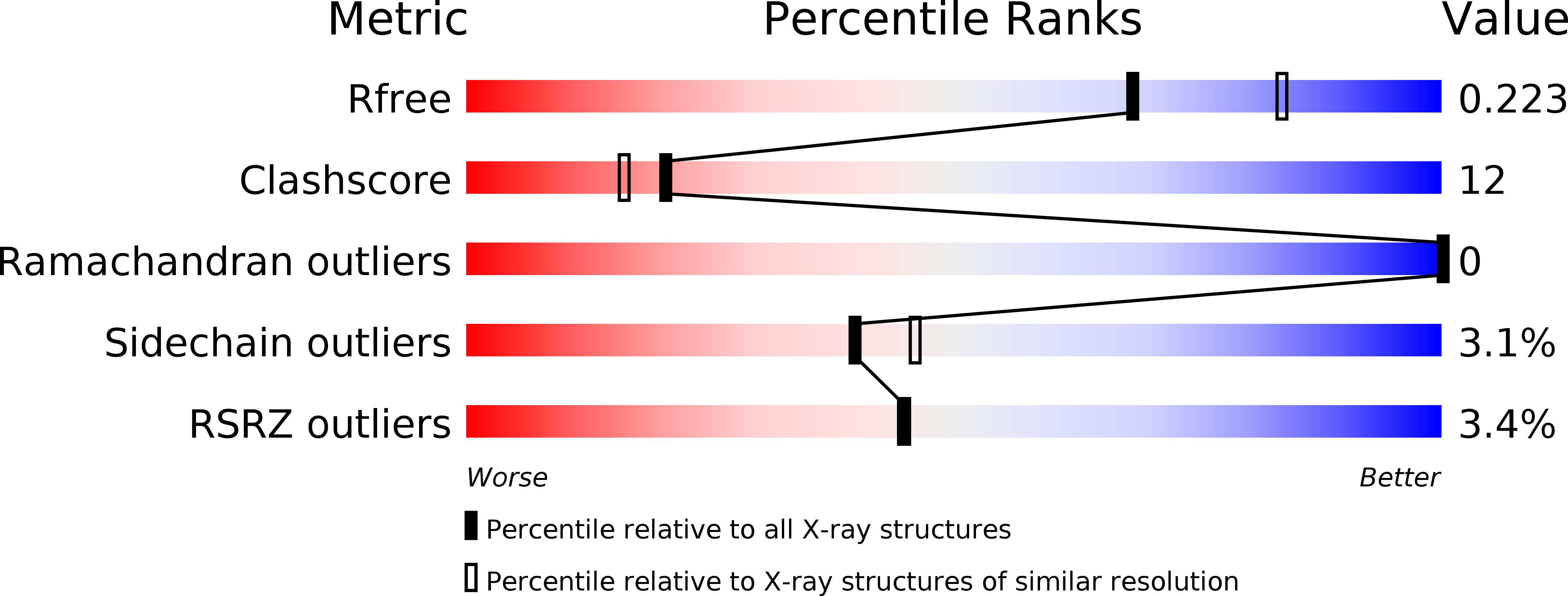
Resolution:
2.24 Å
R-Value Free:
0.22
R-Value Work:
0.19
R-Value Observed:
0.19
Space Group:
P 1 21 1
Macromolecular Entities
Structures with similar UniProt ID
Protein Blast
Polymer Type:polypeptide(L)
Molecule:Aspartate--tRNA ligase, cytoplasmic
Gene (Uniprot):DARS1
Chain IDs:A, B
Chain Length:521
Number of Molecules:2
Biological Source:Homo sapiens
Ligand Molecules
Primary Citation
Crystal structure of human cytosolic aspartyl-tRNA synthetase, a component of multi-tRNA synthetase complex
Proteins 81 1840 1846 (2013)
PMID: 23609930 DOI: 10.1002/prot.24306

Abstact

Human cytosolic aspartyl-tRNA synthetase (DRS) catalyzes the attachment of the amino acid aspartic acid to its cognate tRNA and it is a component of the multi-tRNA synthetase complex (MSC) which has been known to be involved in unexpected signaling pathways. Here, we report the crystal structure of DRS at a resolution of 2.25 Å. DRS is a homodimer with a dimer interface of 3750.5 Å(2) which comprises 16.6% of the monomeric surface area. Our structure reveals the C-terminal end of the N-helix which is considered as a unique addition in DRS, and its conformation further supports the switching model of the N-helix for the transfer of tRNA(Asp) to elongation factor 1α. From our analyses of the crystal structure and post-translational modification of DRS, we suggest that the phosphorylation of Ser146 provokes the separation of DRS from the MSC and provides the binding site for an interaction partner with unforeseen functions.

Legend

Protein

Chemical

Disease

Primary Citation of related structures