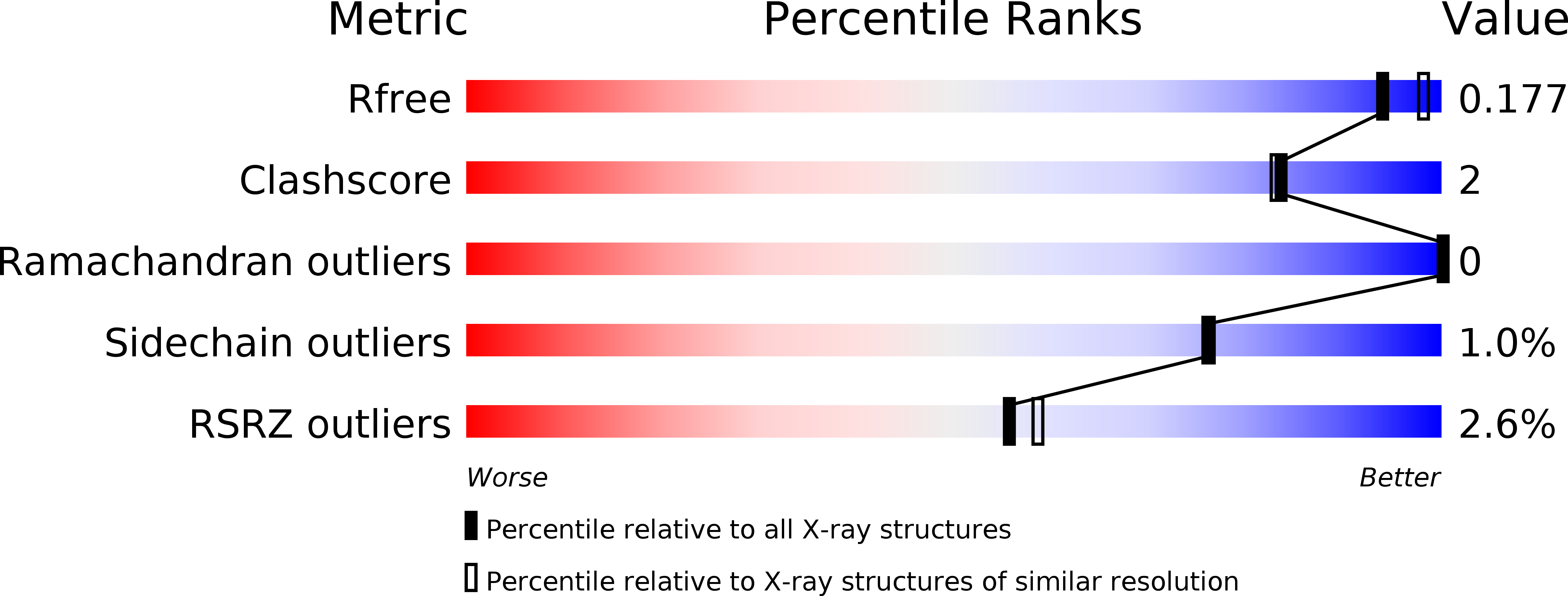
4J09 image
Deposition Date 2013-01-30
Release Date 2014-04-23
Last Version Date 2024-02-28
Entry Detail
PDB ID:
4J09
Keywords:
Title:
Crystal Structure of LpxA bound to RJPXD33
Biological Source:
Source Organism:
Host Organism:
Method Details:
Experimental Method:
Resolution:
1.90 Å
R-Value Free:
0.17
R-Value Work:
0.17
R-Value Observed:
0.17
Space Group:
P 21 3
Macromolecular Entities
Structures with similar UniProt ID
Protein Blast
Polymer Type:polypeptide(L)
Molecule:Acyl-[acyl-carrier-protein]--UDP-N-acetylglucosamine O-acyltransferase
Gene (Uniprot):lpxA
Chain IDs:A
Chain Length:262
Number of Molecules:1
Biological Source:Escherichia coli
Structures with similar UniProt ID
Protein Blast
Polymer Type:polypeptide(L)
Molecule:Putative metabolite transport protein YjhB
Gene (Uniprot):nanX
Chain IDs:B
Chain Length:6
Number of Molecules:1
Biological Source:Escherichia coli
Primary Citation
Structural Basis for the Recognition of Peptide RJPXD33 by Acyltransferases in Lipid A Biosynthesis.
J.Biol.Chem. 289 15527 15535 (2014)
PMID: 24742680 DOI: 10.1074/jbc.M114.564278

Abstact

UDP-N-acetylglucosamine acyltransferase (LpxA) and UDP-3-O-(acyl)-glucosamine acyltransferase (LpxD) constitute the essential, early acyltransferases of lipid A biosynthesis. Recently, an antimicrobial peptide inhibitor, RJPXD33, was identified with dual affinity for LpxA and LpxD. To gain a fundamental understanding of the molecular basis of inhibitor binding, we determined the crystal structure of LpxA from Escherichia coli in complex with RJPXD33 at 1.9 Å resolutions. Our results suggest that the peptide binds in a unique modality that mimics (R)-β-hydroxyacyl pantetheine binding to LpxA and displays how the peptide binds exclusive of the native substrate, acyl-acyl carrier protein. Acyltransferase binding studies with photo-labile RJPXD33 probes and truncations of RJPXD33 validated the structure and provided fundamental insights for future design of small molecule inhibitors. Overlay of the LpxA-RJPXD33 structure with E. coli LpxD identified a complementary peptide binding pocket within LpxD and serves as a model for further biochemical characterization of RJPXD33 binding to LpxD.

Legend

Protein

Chemical

Disease

Primary Citation of related structures