4IZA image
Deposition Date 2013-01-29
Release Date 2013-04-10
Last Version Date 2024-10-30
Entry Detail
PDB ID:
4IZA
Keywords:
Title:
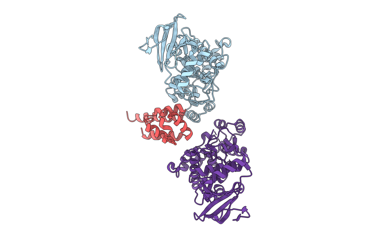
Structure of Dually Phosphorylated ERK2 bound to the PEA-15 Death Effector Domain
Biological Source:
Source Organism:
Homo sapiens (Taxon ID: 9606)
Host Organism:
Method Details:
Experimental Method:
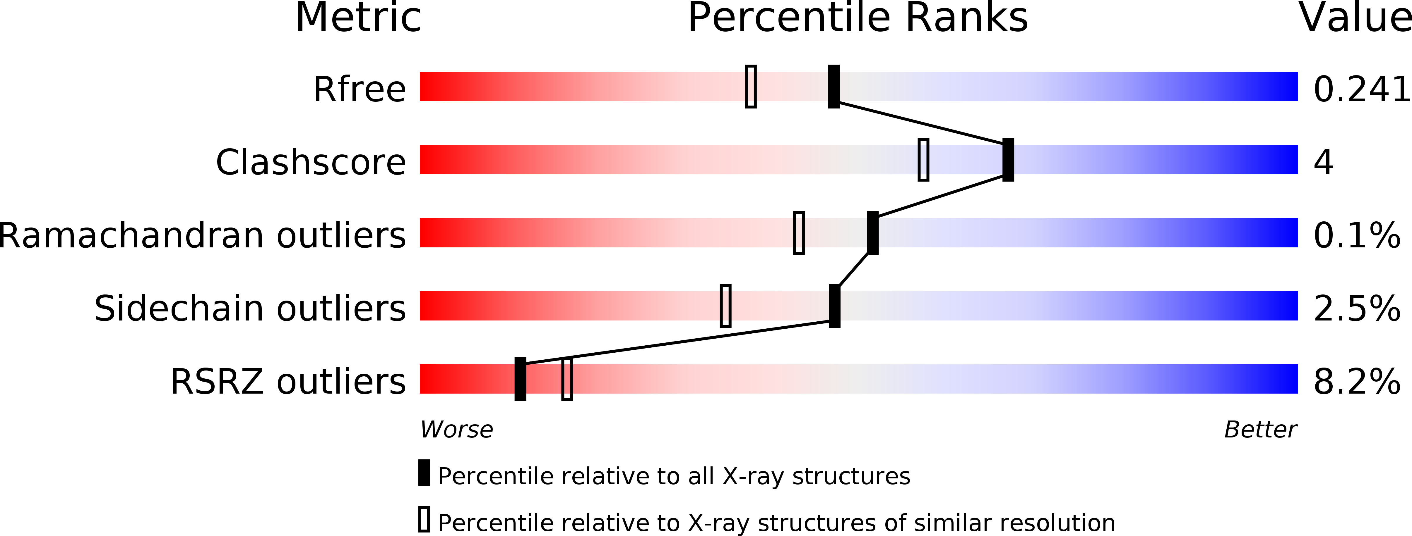
Resolution:
1.93 Å
R-Value Free:
0.24
R-Value Work:
0.19
R-Value Observed:
0.19
Space Group:
P 21 21 2
Macromolecular Entities
Structures with similar UniProt ID
Protein Blast
Polymer Type:polypeptide(L)
Molecule:Mitogen-activated protein kinase 1
Gene (Uniprot):MAPK1
Chain IDs:A, B (auth: C)
Chain Length:356
Number of Molecules:2
Biological Source:Homo sapiens
Structures with similar UniProt ID
Protein Blast
Polymer Type:polypeptide(L)
Molecule:Astrocytic phosphoprotein PEA-15
Gene (Uniprot):PEA15
Chain IDs:C (auth: B)
Chain Length:97
Number of Molecules:1
Biological Source:Homo sapiens
Modified Residue
Compound ID Chain ID Parent Comp ID Details 2D Image
PTR A TYR O-PHOSPHOTYROSINE
TPO A THR PHOSPHOTHREONINE
Primary Citation
Structure of ERK2 bound to PEA-15 reveals a mechanism for rapid release of activated MAPK.
Nat Commun 4 1681 1681 (2013)
PMID: 23575685 DOI: 10.1038/ncomms2687

Abstact

ERK1/2 kinases are the principal effectors of a central signalling cascade that converts extracellular stimuli into cell proliferation and migration responses and, when deregulated, can promote cell oncogenic transformation. The scaffolding protein PEA-15 is a death effector domain protein that directly interacts with ERK1/2 and affects ERK1/2 subcellular localization and phosphorylation. Here, to understand this ERK1/2 signalling complex, we have solved the crystal structures of PEA-15 bound to three different ERK2 phospho-conformers. The structures reveal that PEA-15 uses a bipartite binding mode, occupying two key docking sites of ERK2. Remarkably, PEA-15 can efficiently bind the ERK2 activation loop in the critical Thr-X-Tyr region in different phosphorylation states. PEA-15 binding triggers an extended allosteric conduit in dually phosphorylated ERK2, disrupting key features of active ERK2. At the same time PEA-15 binding protects ERK2 from dephosphorylation, thus setting the stage for immediate ERK activity upon its release from the PEA-15 inhibitory complex.

Legend

Protein

Chemical

Disease

Primary Citation of related structures