4IX9 image
Deposition Date 2013-01-24
Release Date 2013-03-20
Last Version Date 2024-10-16
Entry Detail
PDB ID:
4IX9
Keywords:
Title:
Crystal structure of subunit F of V-ATPase from S. cerevisiae
Biological Source:
Source Organism:
Host Organism:
Method Details:
Experimental Method:
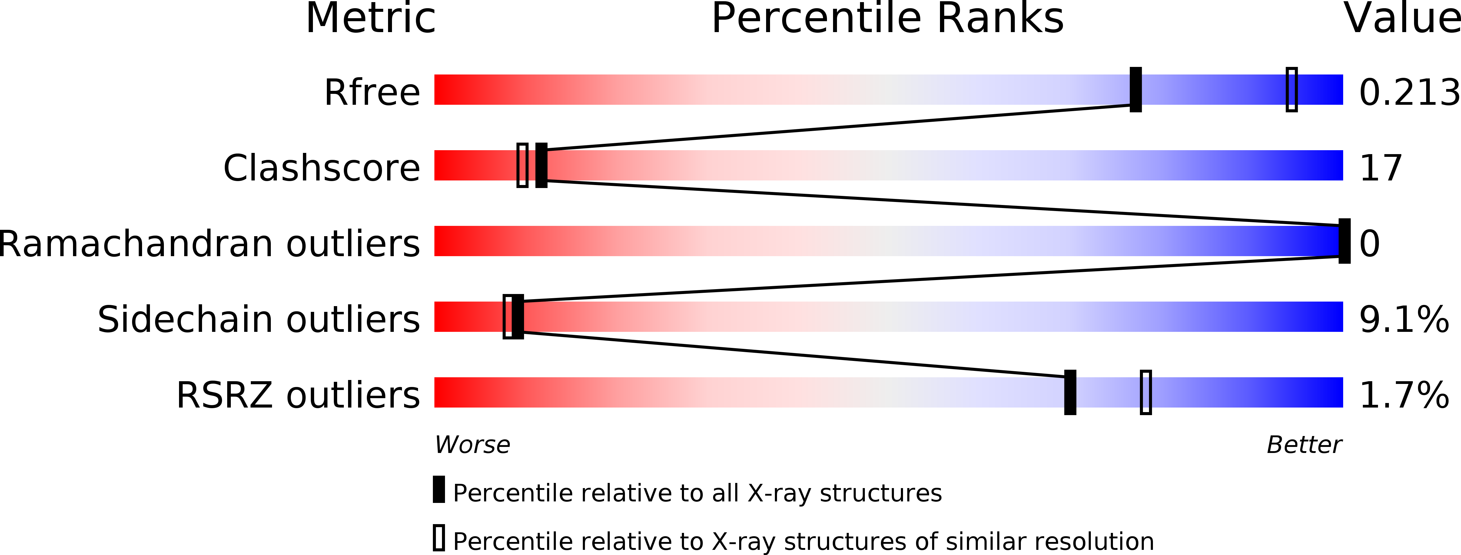
Resolution:
2.33 Å
R-Value Free:
0.21
R-Value Work:
0.15
R-Value Observed:
0.15
Space Group:
C 2 2 21
Macromolecular Entities
Polymer Type:polypeptide(L)
Molecule:V-type proton ATPase subunit F
Gene (Uniprot):VMA7
Mutations:I69M
Chain IDs:A, B, C, D
Chain Length:94
Number of Molecules:4
Biological Source:Saccharomyces cerevisiae
Modified Residue
Compound ID Chain ID Parent Comp ID Details 2D Image
MSE A MET SELENOMETHIONINE
Primary Citation
Crystal and NMR structures give insights into the role and dynamics of subunit F of the eukaryotic V-ATPase from Saccharomyces cerevisiae
J.Biol.Chem. 288 11930 11939 (2013)
PMID: 23476018 DOI: 10.1074/jbc.M113.461533

Abstact

Subunit F of V-ATPases is proposed to undergo structural alterations during catalysis and reversible dissociation from the V1VO complex. Recently, we determined the low resolution structure of F from Saccharomyces cerevisiae V-ATPase, showing an N-terminal egg shape, connected to a C-terminal hook-like segment via a linker region. To understand the mechanistic role of subunit F of S. cerevisiae V-ATPase, composed of 118 amino acids, the crystal structure of the major part of F, F(1-94), was solved at 2.3 Å resolution. The structural features were confirmed by solution NMR spectroscopy using the entire F subunit. The eukaryotic F subunit consists of the N-terminal F(1-94) domain with four-parallel β-strands, which are intermittently surrounded by four α-helices, and the C terminus, including the α5-helix encompassing residues 103 to 113. Two loops (26)GQITPETQEK(35) and (60)ERDDI(64) are described to be essential in mechanistic processes of the V-ATPase enzyme. The (26)GQITPETQEK(35) loop becomes exposed when fitted into the recently determined EM structure of the yeast V1VO-ATPase. A mechanism is proposed in which the (26)GQITPETQEK(35) loop of subunit F and the flexible C-terminal domain of subunit H move in proximity, leading to an inhibitory effect of ATPase activity in V1. Subunits D and F are demonstrated to interact with subunit d. Together with NMR dynamics, the role of subunit F has been discussed in the light of its interactions in the processes of reversible disassembly and ATP hydrolysis of V-ATPases by transmitting movements of subunit d and H of the VO and V1 sector, respectively.

Legend

Protein

Chemical

Disease

Primary Citation of related structures